. 2013 Sep;89(5):949-62.
doi: 10.1111/mmi.12323. Epub 2013 Jul 29.

Substrate specificity of an elongation-specific peptidoglycan endopeptidase and its implications for cell wall architecture and growth of Vibrio cholerae

Affiliations

Substrate specificity of an elongation-specific peptidoglycan endopeptidase and its implications for cell wall architecture and growth of Vibrio cholerae

Tobias Dörr et al. Mol Microbiol. 2013 Sep.

Abstract

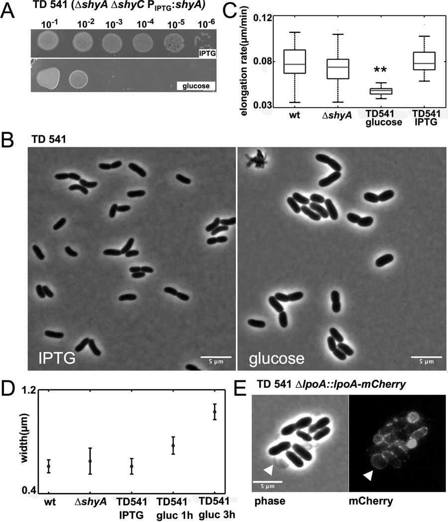
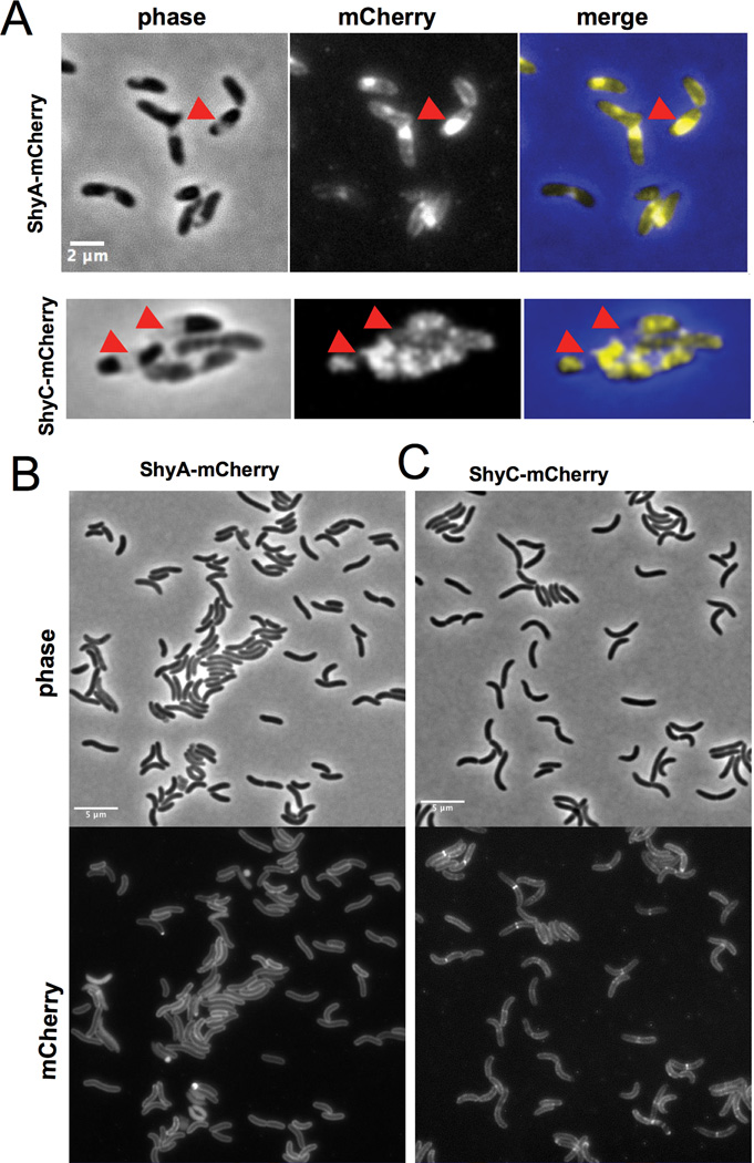
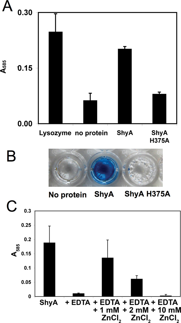
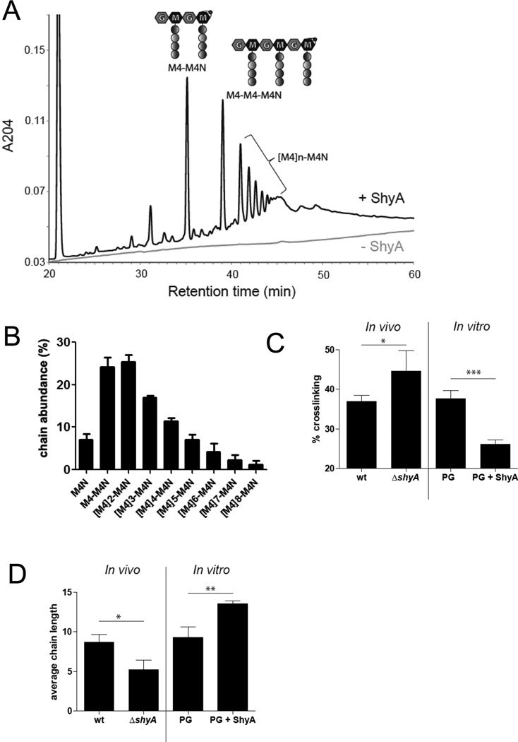
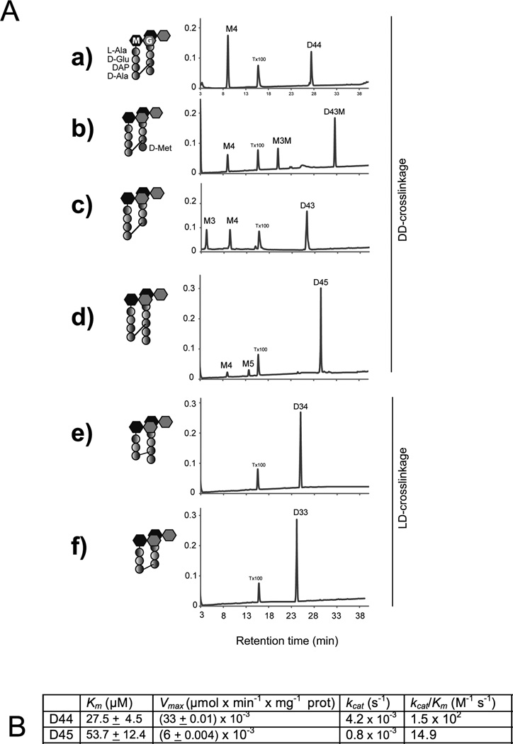
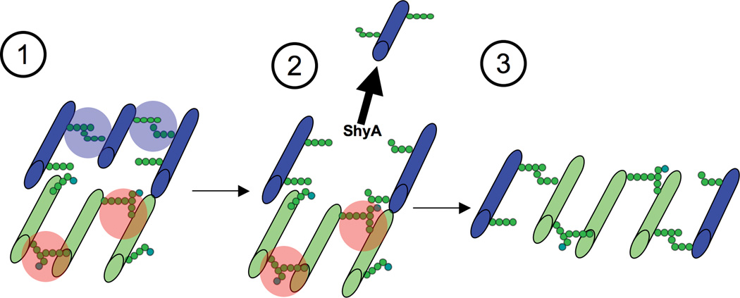
The bacterial cell wall consists of peptidoglycan (PG), a sturdy mesh of glycan strands cross-linked by short peptides. This rigid structure constrains cell shape and size, yet is sufficiently dynamic to accommodate insertion of newly synthesized PG, which was long hypothesized, and recently demonstrated, to require cleavage of the covalent peptide cross-links that couple previously inserted material. Here, we identify several genes in Vibrio cholerae that collectively are required for growth - particularly elongation - of this pathogen. V. cholerae encodes three putative periplasmic proteins, here denoted ShyA, ShyB, and ShyC, that contain both PG binding and M23 family peptidase domains. While none is essential individually, the absence of both ShyA and ShyC results in synthetic lethality, while the absence of ShyA and ShyB causes a significant growth deficiency. ShyA is a D,d-endopeptidase able to cleave most peptide chain cross-links in V. cholerae's PG. PG from a ∆shyA mutant has decreased average chain length, suggesting that ShyA may promote removal of short PG strands. Unexpectedly, ShyA has little activity against muropeptides containing pentapeptides, which typically characterize newly synthesized material. ShyA's substrate-dependent activity may contribute to selection of cleavage sites in PG, whose implications for the process of side-wall growth are discussed.


Figures

Fig. 1
Fig. 1. Growth phenotypes of V. cholerae strains with deletions in orfs containing OapA and M23 peptidase domains
Cells were grown in 200 µL in (A) LB medium or (B) and (C) M9 MM 0.2 % glucose, monitoring OD600. Error bars represent standard deviation of three independent experiments. (D) ShyA depletion in the ΔshyC background. TD541 (ΔshyA ΔshyC PIPTG:shyA) and TD543 (TD541 ΔshyB) were grown in liquid medium in the presence of IPTG, washed, and resuspended in fresh medium containing glucose (T0). Error bars show the standard deviations for three independent experiments.
Fig. 2
Fig. 2. shyA and shyC are synthetically lethal
(A) TD541 (ΔshyA ΔshyC PIPTG:shyA) was grown to stationary phase in LB medium containing IPTG and spotted on LB agar plates containing either IPTG or glucose. (B) ShyA depletion in liquid culture. TD541 was grown in medium containing IPTG, washed twice in LB medium with glucose and resuspended in an equal volume LB/glucose. Cells were imaged just before addition of glucose and three hours after and analyzed using MicrobeTracker. (C) Average elongation rates of around 100 cells of wild type, TD536 (ΔshyA) and TD541 before and after ShyA depletion. Cells were grown on agarose pads with 10 % LB and glucose or IPTG and imaged every minute. Elongation rate is the slope of length increase over time. Asterisks indicate statistical significance (t-test, p<0.01) (D) Average cell widths in liquid cultures of wild type, TD536 and TD541 before and after ShyA depletion. (E) Vesicles emanating from TD541 cells grown on agarose pads contain outer membrane. A functional mCherry fusion of the outer membrane protein LpoA (Vc0581) was expressed from its native promoter and imaged 120 min after plating on an agarose pad containing glucose.
Fig. 3
Fig. 3. ShyA and ShyC have distinct localization patterns
V. cholerae cells expressing mCherry fusions were grown overnight in M9 MM containing 0.2 % glucose and 20 µM IPTG. Cells were then diluted 1:100 into fresh medium containing 100 µM IPTG and grown for 2 h, applied to a 0.8 % agarose pad containing M9 MM glucose and imaged with epifluorescence (500 ms exposure). (A) Cells imaged after 15 min exposure to 15 % sucrose (see experimental procedures for details). Arrowheads point to enlarged periplasmic pockets that result from withdrawal of the inner membrane. (B) and (C) Phase contrast and fluorescence images of V. cholerae carrying (B) pshyA-mCherry and (C) pshyC mCherry.
Fig. 4
Fig. 4. ShyA has PG hydrolase activity in vitro
(A) Remazol-Blue (RBB) stained sacculi were incubated with 5 µM of purified ShyA-His, ShyAH375A-His, or lysozyme (a positive control) for 3 h at 37˙C. Undigested material was removed by ultracentrifugation, then OD585 of the supernatant was measured. Error bars represent standard deviations of 3 independent experiments. (B) Representative picture of supernatants from mock or ShyA-digested RBB-stained sacculi after ultracentifugation. (C) RBB stained sacculi were treated as described in (A) with the additions indicated on the x-axis. Error bars represent standard deviations of two independent experiments.
Fig. 5
Fig. 5. ShyA is a D,D-endopeptidase
(A) Representative HPLC chromatograms of the supernatant of undigested sacculi (lower line) and sacculi digested with ShyA (peaks). PG was digested with 5 µM ShyA for 3 h at 37°C. (B) Relative abundance of PG-chains released by ShyA. Sacculi were digested with ShyA as in (A) and PG chains were quantified using HPLC (see experimental procedures for details). (C) Quantification of PG crosslinks in sacculi from wild type (wt) and ΔshyA cells (In vivo) as well as of wild type PG partially digested (0.1 µM for 1 h at ambient temperature) with ShyA (PG+ ShyA) or undigested (PG) (in vitro). P values (wt vs ΔshyA): 0.0447; (PG vs PG + ShyA): 0.0010. (D) Average chain length of the wt and ΔshyA mutant as well as PG partially digested with ShyA as in (B). Chain length was estimated based on the relative abundance of anhydromuropeptides. P values (wt vs ΔshyA): 0.0181; (PG vs PG + ShyA): 0.0061. (B–D) correspond to three independent experiments each, error bars represent standard deviation.
Fig. 6
Fig. 6. ShyA substrate preferences
(A) HPLC analysis of ShyA endopeptidase activities on muropeptides The indicated DD-crosslinked dimers (D44, D43, D43M, D45) and LD-crosslinked dimer muropeptide substrates (D33 and D34) were incubated with 7.8 µM ShyA-His for 3h at 37˙C before HPLC analysis. ShyA endopeptidase activity was assayed by monitoring the appearance of the monomeric compounds M4 (GM-L-Ala-D-Glu-DAP-D-Ala); M3 (GlcNAc-MurNAc-L-Ala-D-Glu-DAP), M5 (GM-L-Ala-D-Glu-DAP-D-Ala-D-Ala) and M3M (GM-L-Ala-D-Glu-DAP-D-Met). Assays were carried out in duplicates and representative HPLC chromatograms are shown. X-axis = A204 (B) Kinetics of ShyA-His D,D-endopeptidase activities. D,D-endopeptidase activity in vitro on D44 and D45 muropeptide substrates was measured as described in experimental procedures. All kinetic constants were calculated using data obtained with 7.8 µM ShyA with various amounts (1–300 µM) of the dimers bis-dissacharide tetratetrapeptide (D44) and bis-dissacharide tetrapentapeptide (D45). The enzymatic reactions were analyzed by HPLC assay as described in experimental procedures. All kinetic constants must be considered apparent values because of the impossibility of calculating initial enzyme velocities by HPLC. Values are means ± standard deviations of experimental triplicates.
Fig. 7
Fig. 7. Model of ShyA-mediated sacculus enlargement
(1) New PG (colored green) is synthesized and contains ShyA-resistant 4/5 peptide bonds (shaded red, the fifth D-Ala is colored light blue). Old PG (colored blue) contains mostly ShyA-sensitive 4/4 crosslinks (shaded blue). We follow the “three for one” model proposed by Höltje (Holtje, 1998) and thus assume that three new strands are synthesized for incorporation. (2) ShyA cleaves and removes an old PG strand without affecting the bonds in new PG. Newly synthesized PG might either be already connected to the old material (as originally proposed in the three for one model) or it might be crosslinked concomitant with ShyA action. (3) Three nascent strands replace one old strand, enlarging the PG mesh.

References

    1. Bisicchia P, Noone D, Lioliou E, Howell A, Quigley S, Jensen T, Jarmer H, Devine KM. The essential YycFG two-component system controls cell wall metabolism in Bacillus subtilis . Mol Microbiol. 2007;65:180–200. - PubMed
    1. Butterton JR, Beattie DT, Gardel CL, Carroll PA, Hyman T, Killeen KP, Mekalanos JJ, Calderwood SB. Heterologous antigen expression in Vibrio cholerae vector strains. Infect Immun. 1995;63:2689–2696. - PMC - PubMed
    1. Cabeen MT, Jacobs-Wagner C. Bacterial cell shape. Nat Rev Microbiol. 2005;3:601–610. - PubMed
    1. Cava F, de Pedro MA, Lam H, Davis BM, Waldor MK. Distinct pathways for modification of the bacterial cell wall by non-canonical D-amino acids. EMBO J. 2010;30:3442–3453. - PMC - PubMed
    1. Chowdhury C, Nayak TR, Young KD, Ghosh AS. A weak DD-carboxypeptidase activity explains the inability of PBP 6 to substitute for PBP 5 in maintaining normal cell shape in Escherichia coli . FEMS Microbiol Lett. 2010;303:76–83. - PMC - PubMed

Publication types

MeSH terms

LinkOut - more resources