. 2013 Sep;13(9):2268-79.
doi: 10.1111/ajt.12342. Epub 2013 Jul 8.

Transient lymphopenia breaks costimulatory blockade-based peripheral tolerance and initiates cardiac allograft rejection

Affiliations

Transient lymphopenia breaks costimulatory blockade-based peripheral tolerance and initiates cardiac allograft rejection

S Iida et al. Am J Transplant. 2013 Sep.

Abstract

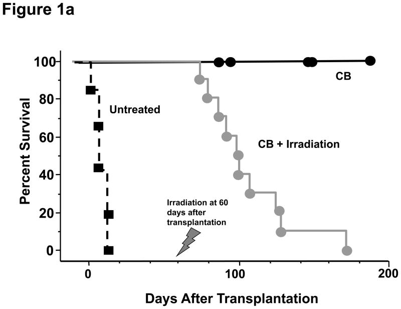
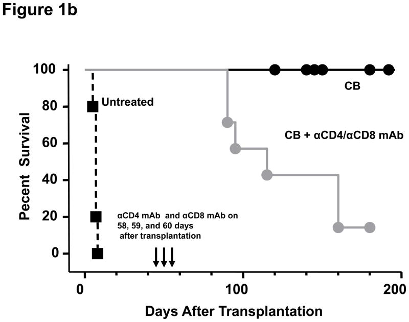
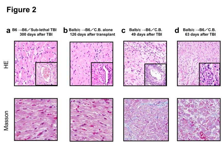
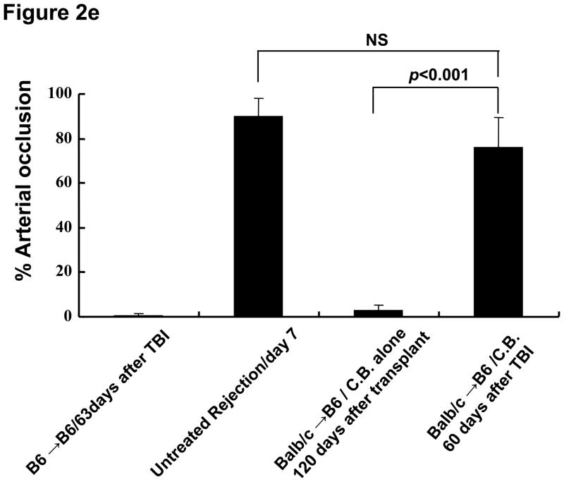
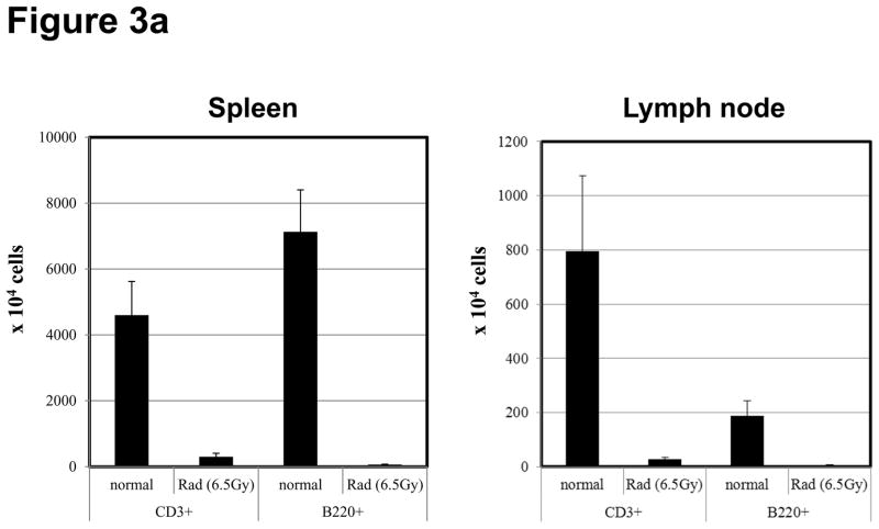
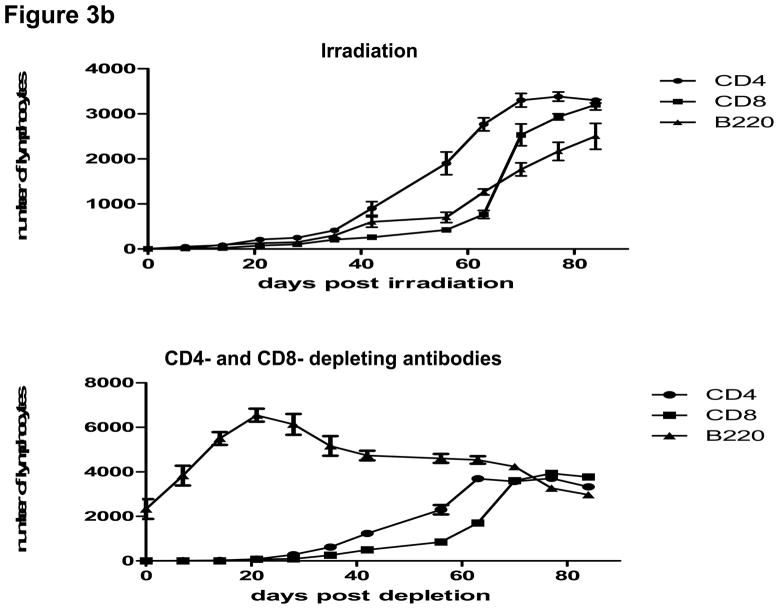
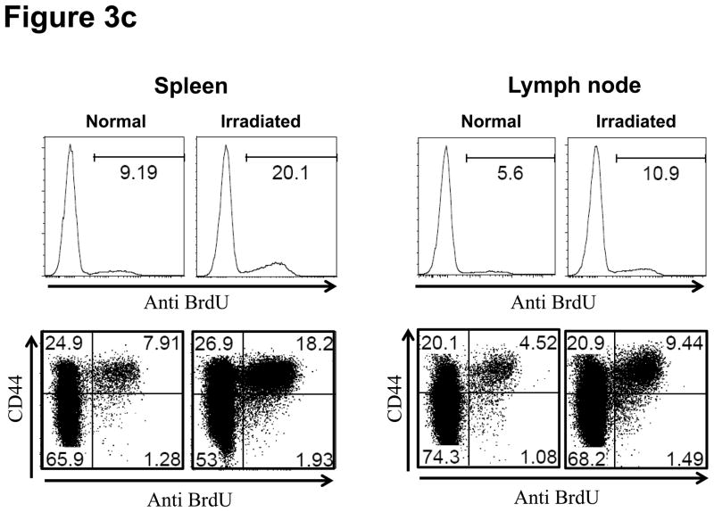
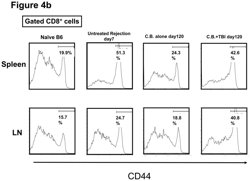
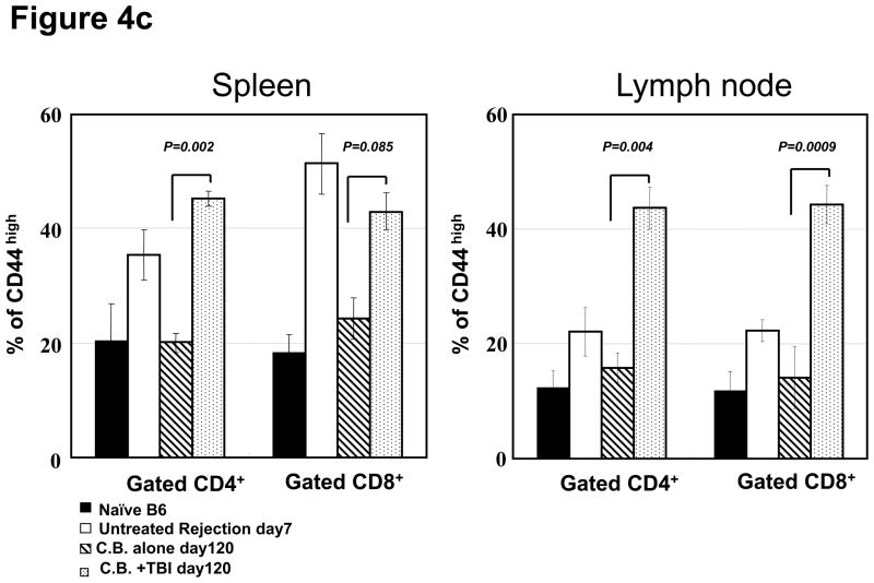
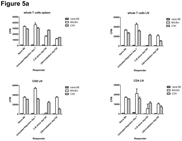
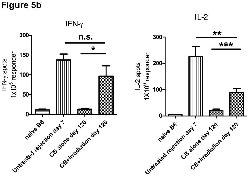
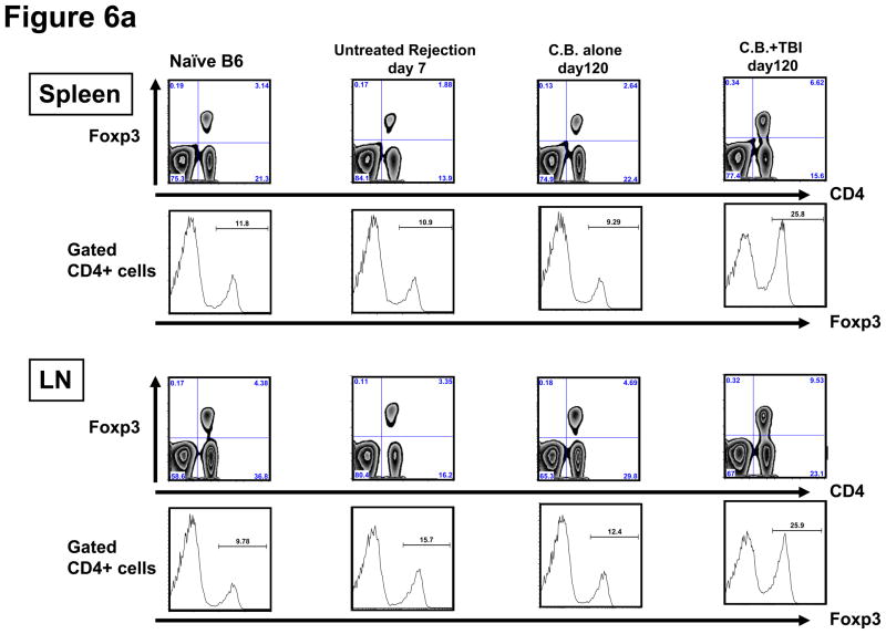
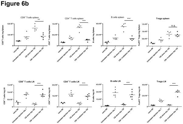
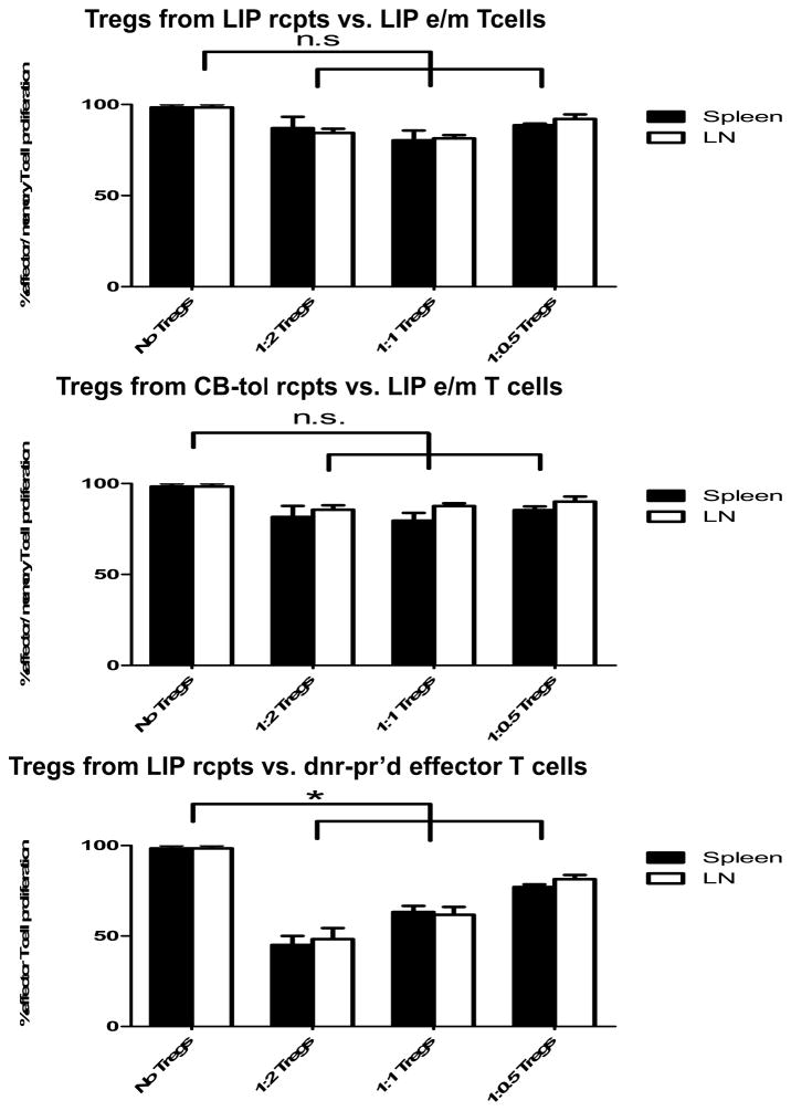
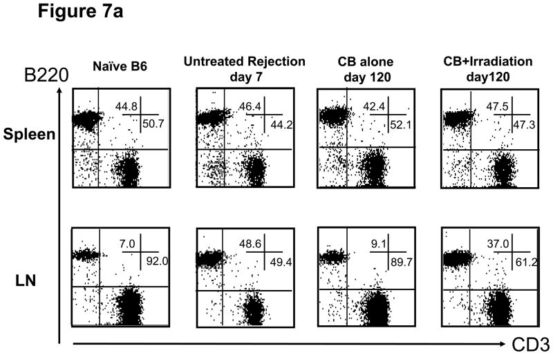
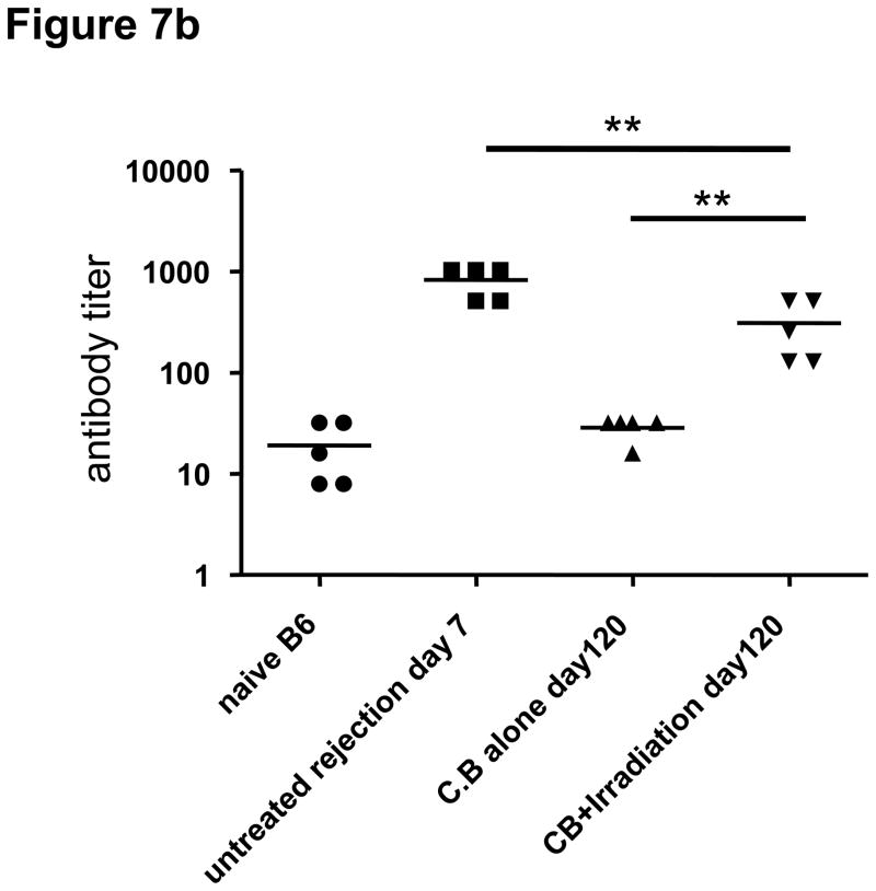
Lymphopenia is induced by lymphoablative therapies and chronic viral infections. We assessed the impact of lymphopenia on cardiac allograft survival in recipients conditioned with peritransplant costimulatory blockade (CB) to promote long-term graft acceptance. After vascularized MHC-mismatched heterotopic heart grafts were stably accepted through CB, lymphopenia was induced on day 60 posttransplant by 6.5 Gy irradiation or by administration of anti-CD4 plus anti-CD8 mAb. Long-term surviving allografts were gradually rejected after lymphodepletion (MST = 74 ± 5 days postirradiation). Histological analyses indicated signs of severe rejection in allografts following lymphodepletion, including mononuclear cell infiltration and obliterative vasculopathy. Lymphodepletion of CB conditioned recipients induced increases in CD44(high) effector/memory T cells in lymphatic organs and strong recovery of donor-reactive T cell responses, indicating lymphopenia-induced proliferation (LIP) and donor alloimmune responses occurring in the host. T regulatory (CD4(+) Foxp(3+)) cell and B cell numbers as well as donor-specific antibody titers also increased during allograft rejection in CB conditioned recipients given lymphodepletion. These observations suggest that allograft rejection following partial lymphocyte depletion is mediated by LIP of donor-reactive memory T cells. As lymphopenia may cause unexpected rejection of stable allografts, adequate strategies must be developed to control T cell proliferation and differentiation during lymphopenia.

Keywords: Costimulatory blockade; lymphopenia-induced proliferation; memory T cell.


Conflict of interest statement

Disclosures

The authors have no financial conflict of interest to declare.

Figures

Figure 1
Figure 1. Transient lymphopenia leads to rejection of cardiac allografts in CB conditioned recipients
C57BL/6 mice received complete MHC-mismatched BALB/c heterotopic heart transplants with or without 500 μg of anti-CD40L mAb plus 500 μg of hCTLA4-Ig (CB conditioning) on the day of transplantation and every other day for 6 days to promote long-term allograft survival. One group of the CB conditioned recipients was (a) sub-lethally irradiated (6.5 Gy on day 60 post-transplant; and, (b) another group was treated on days 58, 59, and 60 post-transplant with CD4− and CD8− depleting antibodies. Allograft survival was monitored by palpation.
Figure 2
Figure 2. Histological examination of representative cardiac grafts in irradiated hosts
a–d) Cardiac grafts were harvested from B6 recipients at the indicated time points post-transplant and prepared sections stained with H&E (upper row) or Masson’s trichrome (lower row). Original magnification ×200; inserts ×400. e) Bars represent the combined mean ± SEM of the occlusion area of heart grafts observed in 10–20 microscopic frames of view taken from no fewer than six different cardiac grafts per group.
Figure 3
Figure 3. Lymphopenia and subsequent lymphopenia-induced proliferation of residual T cells following lymphocytes depletion
a) Naive C57BL/6 mice were subjected to 6.5Gy whole body irradiation and numbers of T and B cells in the spleen and LN were assessed 5 days later by antibody staining and flow cytometry analysis. b) The kinetics of lymphocyte population recovery in the peripheral blood of CB-conditioned heart allograft recipients following irradiation or treatment with CD4- plus CD8-depleting mAb as assessed by antibody staining and flow cytometry analysis. c) The proliferation of residual T cells in the spleen and LN 30 days of naïve mice after irradiation as assessed by BrdU incorporation.
Figure 4
Figure 4. Appearance of CD4 and CD8 T cells expressing high levels of CD44 following transient lymphopenia
a and b) The presence of CD44high, memory phenotype CD4+ and CD8+ T cells in the spleen and LN of naïve C57BL/6 mice, untreated C57BL/6 recipients of BALB/c cardiac allografts on day 7 post-transplant, CB-conditioned, tolerant allograft recipients on day 120 post-transplant, and CB-conditioned allograft recipients subjected to irradiation on day 120 post-transplant. c) The frequency of CD44high T cells among total CD4+ or CD8+ T cells in the spleen and LN of the mice in analyzed in a and b. Data are expressed as mean % CD44high in gated CD4+ and CD8+ T cell populations ± SD. P values were calculated using the two-tailed paired Students’ t-test.
Figure 5
Figure 5. Donor-reactive T cells responses are recovered in CB-conditioned heart allograft recipients following transient lymphopenia
a) Spleen and LN cell suspensions were prepared from naïve C57BL/6 mice, untreated C57BL/6 recipients of BALB/c cardiac allografts on day 7 post-transplant, CB-conditioned, tolerant allograft recipients on day 120 post-transplant, and CB-conditioned allograft recipients subjected to irradiation on day 120 post-transplant. As indicated unseparated cells or enriched CD4+ or CD8+ T cell populations were cultured with syngeneic, BALB/c donor-derived, or third-party allogeneic C3H spleen cells that had been depleted of T cells for 88 hours and then pulsed with 3H-thymidinde for 8 hours before harvest and assessment of 3H-thymidine incorporation. Data represent the mean ± SEM 3H-thymidine incorporation of triplicate cultures from a single experiment that is representative of three others. b) T cells were enriched from LNs by negative selection and were cultured with BALB/c donor spleen cells that had been depleted of T cells to enumerate donor-reactive T cells producing IFN-γ and IL-2 by ELISPOT assay. The data indicate the mean number of donor-reactive T cells producing the test cytokine ± SEM. *p = 0.032 versus CB-tolerant recipients; **p = 0.014 versus CB-tolerant recipients; ***p = 0.029 versus untreated rejection recipients. Data are representative of two independent experiments.
Figure 6
Figure 6. Transient lymphopenia of CB-conditioned allograft recipients increases Foxp3+ CD4+ T cells in LN and spleen without suppressing the alloreactive effector/memory T cell response
a) Spleen and LN cell suspensions were prepared from naïve C57BL/6 mice, untreated C57BL/6 recipients of BALB/c cardiac allografts on day 7 post-transplant, CB-conditioned, tolerant allograft recipients on day 120 post-transplant, and CB-conditioned allograft recipients subjected to irradiation on day 120 post-transplant and stained with anti-CD4 and anti-FoxP3 mAb to determine the percentage of double positive cells. The percentage of Foxp3+ cells in the CD4+ T cell population was higher in irradiated recipients than in the other groups (p < 0.001, for both spleen and LN). b) The total numbers of CD8 T, CD4 T, B cells, and CD4+FoxP3+ Treg cells per mg of lymphoid tissue was determined by antibody staining and flow cytometry analysis at the time of spleen and LN harvest of each group. **p < 0.01; ***p < 0.001. c) For test Treg cells, CD4+CD25+ T cells were isolated using magnetic beads (Stem Cell Technologies, Vancouver, BC) on day 120 post-transplant from the spleen and LN of either CB-conditioned, tolerant allograft recipients or CB-conditioned allograft recipients subjected to irradiation. For use as LIP effector/memory responder T cells, T cells from LIP mice on day 120 post-transplant were flow sorted to isolate CD62Llow, CD44high T cells (> 98% purity) using a FACSAria (BD Biosciences, San Jose, CA). For use as primary effector T cell responders, T cells from the spleen and lymph nodes of LIP mice were obtained on day 120 post-transplant and the CD4+CD25+ T cells removed. Responder T cell populations were labeled with CFSE and these cells and the isolated Treg cell populations added to cultures of donor-derived T cell depleted spleen cells and anti-CD3 mAb in the indicated responder T cell: regulatory T cell ratios. After 4 days of culture, the degree of responder T cell proliferation was determined by assessing CFSE dilution in flow cytometry analyses. All cultures were performed and analyzed in triplicate, and levels of proliferation in the absence of Tregs was set at 100%. n.s., p > 0.05.
Figure 7
Figure 7. Transient lymphopenia in CB conditioned allograft recipients induces the accumulation of B cells in lymph nodes and restores antibody production against allogenic antigens
a) Flow cytometry analysis of T and B cells in the spleen and LN of naïve C57BL/6 mice, untreated C57BL/6 recipients of BALB/c cardiac allografts on day 7 post-transplant, CB-conditioned, tolerant allograft recipients on day 120 post-transplant, and CB-conditioned allograft recipients subjected to irradiation on day on day 120 post-transplant. b) Serum from naïve, untreated-rejecting allograft recipients on day 7 post-transplant, CB-conditioned, tolerant allograft recipients on day 120 post-transplant, and CB-conditioned allograft recipients subjected to irradiation on day on day 120 post-transplant was collected and tested for reactivity to BALB/c thymocytes using flow cytometry-based analyses to determine donor-reactive antibody titers. Data indicate mean titer ± SEM for n=5/group and are representative of two independent experiments. **p < 0.01

Similar articles

Cited by

References

    1. Baczkowska T, Durlik M. Calcineurin inhibitor sparing immunosuppressive regimens in kidney allograft recipients. Pol Arch Med Wewn. 2009 May;119(5):318–25. - PubMed
    1. Vathsala A. Preventing renal transplant failure. Ann Acad Med Singapore. 2005 Jan;34(1):36–43. - PubMed
    1. Boisgerault F, Liu Y, Anosova N, Ehrlich E, Dana MR, Benichou G. Role of CD4+ and CD8+ T cells in allorecognition: lessons from corneal transplantation. J Immunol. 2001 Aug 15;167(4):1891–9. - PubMed
    1. Baldwin WM, 3rd, Valujskikh A, Fairchild RL. Antibody-mediated rejection: emergence of animal models to answer clinical questions. Am J Transplant. May;10(5):1135–42. - PMC - PubMed
    1. Billing H, Rieger S, Ovens J, Susal C, Melk A, Waldherr R, et al. Successful treatment of chronic antibody-mediated rejection with IVIG and rituximab in pediatric renal transplant recipients. Transplantation. 2008 Nov 15;86(9):1214–21. - PubMed

Publication types