

Abdominal cerebrospinal fluid pseudocyst occurring 21 years after ventriculoperitoneal shunt placement: a case report
- PMID: 23834856
- PMCID: PMC3710075
- DOI: 10.1186/1471-2482-13-27
Abdominal cerebrospinal fluid pseudocyst occurring 21 years after ventriculoperitoneal shunt placement: a case report
Abstract
Background: Ventriculoperitoneal shunt (VPS) placement is an established procedure for the treatment of hydrocephalus of diverse etiologies in children and adults. Abdominal cerebrospinal fluid pseudocyst, which is potentially life threatening, is a rare complication and usually occurs during childhood. However, with increasing longevity following successful treatment, it can also occur in adults.
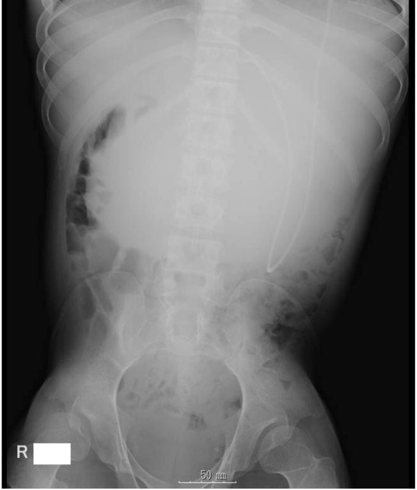
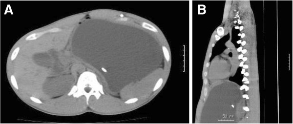
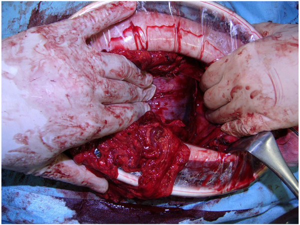
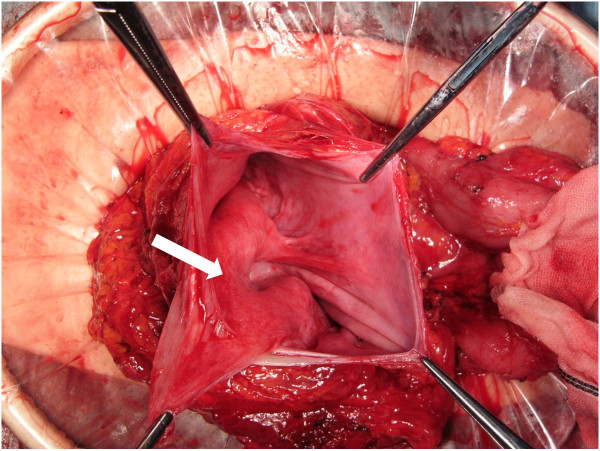
Case presentation: Here we describe a 22-year-old man who was admitted to our hospital because of diffuse abdominal distention. A VPS was placed 21 years earlier to treat hydrocephalus secondary to spina bifida. Abdominal computed tomography (CT) revealed a homogeneous low-density fluid collection adjacent to the VPS catheter tip, causing stomach obstruction. Thus a peritoneal pseudocyst around VPS was suspected and emergency laparotomy was performed. The large mass was localized in the left upper abdomen between the stomach and mesentery of the transverse colon, exactly at the omental bursa. The cystic mass was opened and 1500 ml of clear fluid was drained; the distal end of the VPS was repositioned outside the mass. Thus, an abdominal cerebrospinal fluid pseudocyst as a complication of VPS was diagnosed.
Conclusion: Gastroenterological surgeons should be aware of this possible complication, and this complication should be considered during differential diagnosis of an acute abdomen complaint.
Figures
References
-
- Sena FG, Sousa RM, Meguins LC. Abdominal cerebrospinal fluid pseudocyst: a complication of ventriculoperitoneal shunt in a Brazilian Amazon woman. Case report. Il Giorn Chir. 2010;31(8–9):371–373. - PubMed
-
- Oh A, Wildbrett P, Golub R, Yu LM, Goodrich J, Lee T. Laparoscopic repositioning of a ventriculo-peritoneal catheter tip for a sterile abdominal cerebrospinal fluid (CSF) pseudocyst. Surg Endosc. 2001;15(5):518. - PubMed
Publication types
MeSH terms
LinkOut - more resources
Full Text Sources
Other Literature Sources
