. 2013 Jul 8:11:78.
doi: 10.1186/1741-7007-11-78.

Ascl1b and Neurod1, instead of Neurog3, control pancreatic endocrine cell fate in zebrafish

Affiliations

Ascl1b and Neurod1, instead of Neurog3, control pancreatic endocrine cell fate in zebrafish

Lydie C Flasse et al. BMC Biol. .

Abstract

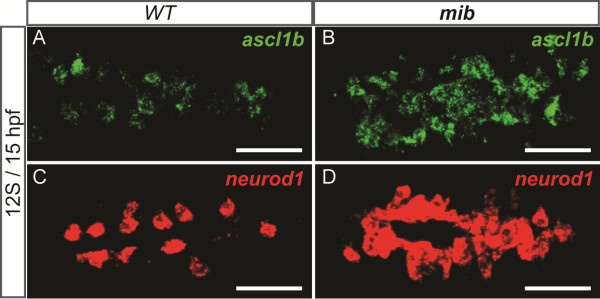
Background: NEUROG3 is a key regulator of pancreatic endocrine cell differentiation in mouse, essential for the generation of all mature hormone producing cells. It is repressed by Notch signaling that prevents pancreatic cell differentiation by maintaining precursors in an undifferentiated state.

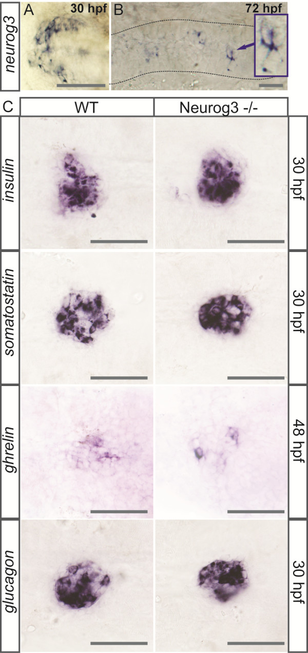
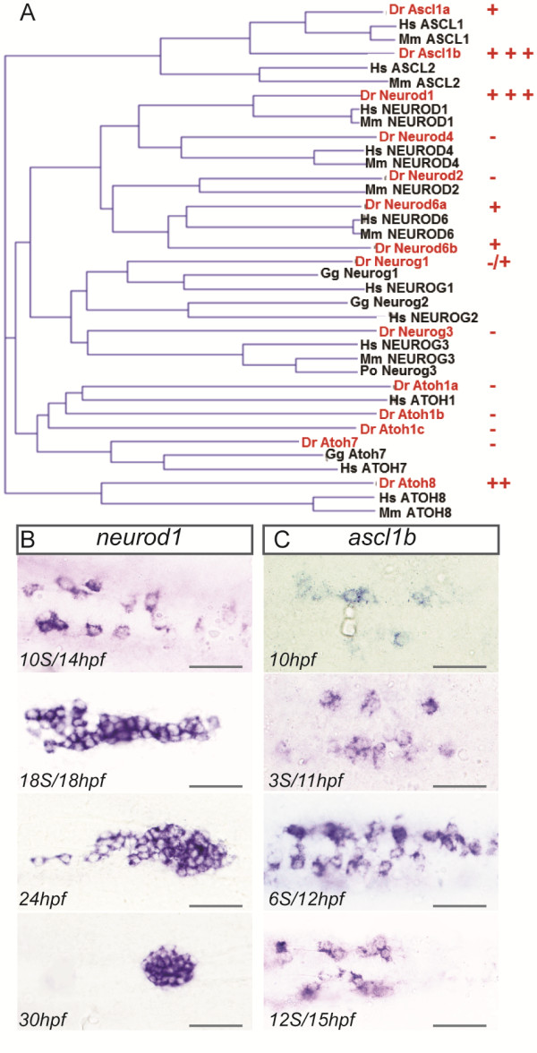
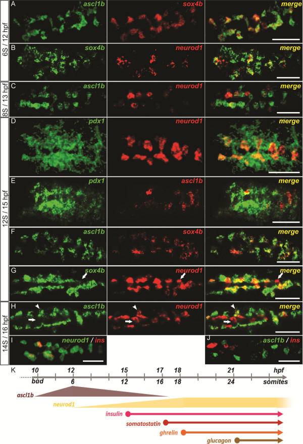
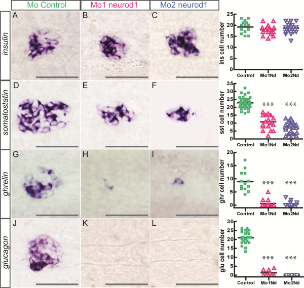
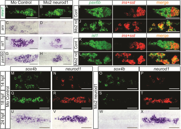
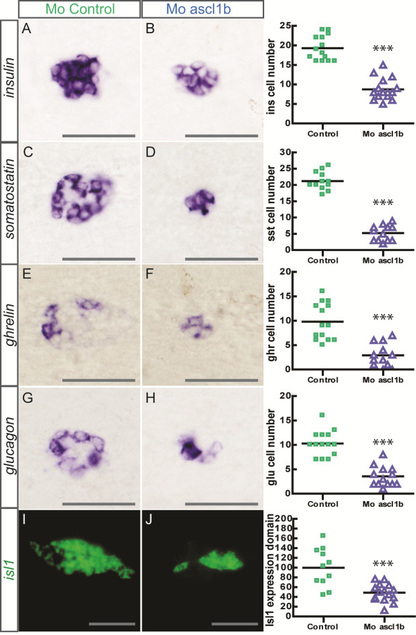
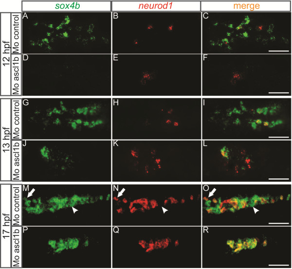
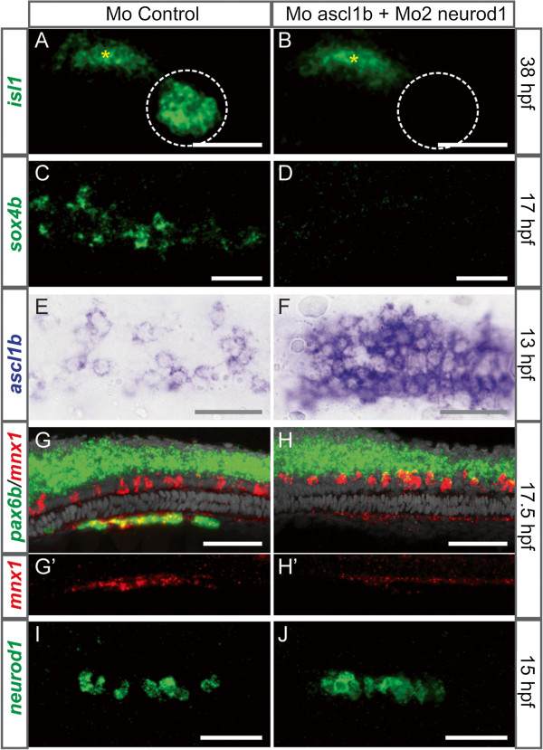
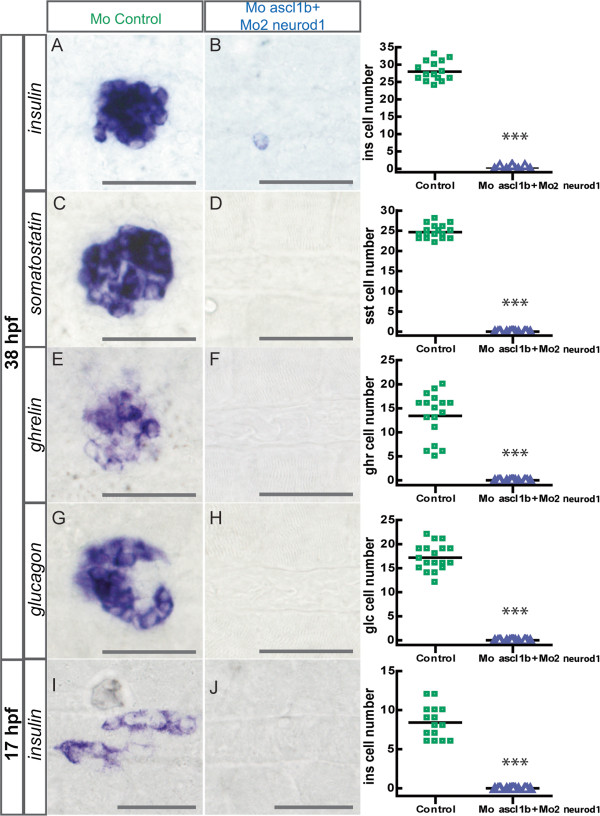
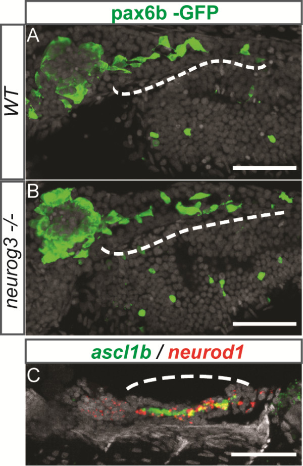
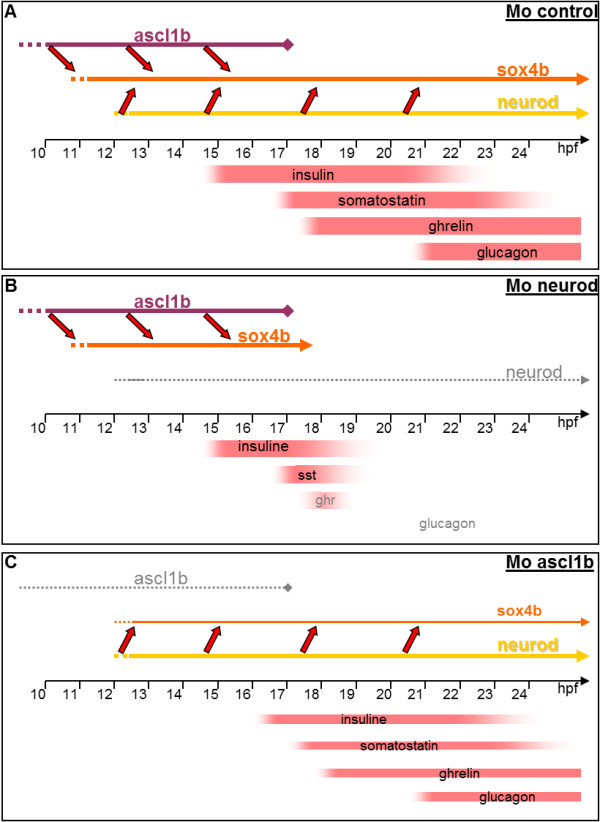
Results: We show that, in zebrafish, neurog3 is not expressed in the pancreas and null neurog3 mutant embryos do not display any apparent endocrine defects. The control of endocrine cell fate is instead fulfilled by two basic helix-loop-helix factors, Ascl1b and Neurod1, that are both repressed by Notch signaling. ascl1b is transiently expressed in the mid-trunk endoderm just after gastrulation and is required for the generation of the first pancreatic endocrine precursor cells. Neurod1 is expressed afterwards in the pancreatic anlagen and pursues the endocrine cell differentiation program initiated by Ascl1b. Their complementary role in endocrine differentiation of the dorsal bud is demonstrated by the loss of all hormone-secreting cells following their simultaneous inactivation. This defect is due to a blockage of the initiation of endocrine cell differentiation.

Conclusions: This study demonstrates that NEUROG3 is not the unique pancreatic endocrine cell fate determinant in vertebrates. A general survey of endocrine cell fate determinants in the whole digestive system among vertebrates indicates that they all belong to the ARP/ASCL family but not necessarily to the Neurog3 subfamily. The identity of the ARP/ASCL factor involved depends not only on the organ but also on the species. One could, therefore, consider differentiating stem cells into insulin-producing cells without the involvement of NEUROG3 but via another ARP/ASCL factor.


Figures

Figure 1
Figure 1
neurog3 is not expressed in the pancreas and null neurog3 mutant embryos do not display any apparent endocrine defects. (A-B) Whole-mount in situ hybridization (WISH) showing expression of neurog3 in the hypothalamus at 30 hpf (A) and in scattered cells of the gut (dotted lines) at 72 hpf (B). (C) WISH showing that the number of cells expressing insulin, somatostatin, ghrelin and glucagon is not changed in the neurog3 sa211 mutants compared to the wt siblings. All views are ventral with the anterior part to the left. Scale bars : 50 µm hpf, hours post fertilization; wt, wild type.
Figure 2
Figure 2
ascl1b and neurod1 are expressed during early pancreas development. (A) Phylogenetic tree calculated with full-length amino acid sequences from vertebrate members of the ARP/ASCL family. The pancreatic expression of zebrafish ARP/ASCL was tested by WISH and the expression levels are depicted by (+++) highly, (++) moderately, (+) weakly, (-/+) very weakly and (-) not expressed in the pancreas. (B-C) WISH showing expression of neurod1(B) and ascl1b(C) in the pancreatic region at the indicated stages. All views are ventral with anterior part to the left. Scale bars : 50 µm WISH, whole-mount in situ hybridization.
Figure 3
Figure 3
ascl1b and neurod1 are both expressed in pancreatic endocrine precursors. (A-J) Double fluorescent WISH comparing the expression of ascl1b and neurod1 with the expression of the pancreatic factor sox4b(A, B, F, G), pdx1(D, E) or with the insulin hormone (I, J) at early stages (12 to 16 hpf). The arrows point to a cell neurod1+/sox4b- (G) or neurod1+/ascl1b- (H). The arrowheads point to a cell neurod1+/ascl1b+(H). Z-plane confocal images. All views are ventral with anterior part to the left. (K) Diagram illustrating the time windows for the expression of ascl1b, neurod1 and the pancreatic hormones. Scale bars : 50 µm hpf, hours post fertilization; WISH, whole-mount in situ hybridization.
Figure 4
Figure 4
Pancreatic expression of ascl1b and neurod1 is repressed by Notch signaling. Fluorescent WISH showing pancreatic expression of ascl1b(A-B) and neurod1(C-D) in wild-type (wt) embryos (A, C) and mind bomb (mib) mutants (B, D). Confocal projection images. All views are ventral views of 15 hpf embryos with anterior part to the left. Scale bars : 50 µm hpf, hours post fertilization; WISH, whole-mount in situ hybridization.
Figure 5
Figure 5
Impaired endocrine cell differentiation in neurod1 morphants. Ventral views with anterior to the left of WISH showing expression of insulin(A-C), somatostatin(D-F), ghrelin(G-I) and glucagon(J-L) in control (A, D, G, J), Mo1-neurod1(B, E, H, K) and Mo2-neurod1(C, F, I, L) morphants at 30 hpf. The quantifications on the right side of the figure represent the number of positive cells per embryo for controls and neurod1 morphants. Asterisks (***) indicate that the difference between cell number in controls and neurod1 morphants is statistically significant by Student’s t-test (P <0.001). Scale bars : 50 µm hpf, hours post fertilization; WISH, whole-mount in situ hybridization.
Figure 6
Figure 6
Pancreatic cells are arrested in their differentiation process in neurod1 morphants and the expression of sox4b is not maintained. Ventral views with the anterior to the left of the pancreas of embryos analyzed by visible (C-H, U-X) or fluorescent (A-B, I-T) WISH. (A-H) Pancreatic expression of mnx1, arx, isl1 and pax6b in controls and Mo2-neurod1 morphants at 24 hpf, the number of isl1+ cells in control morphant (50.4 ± 2.1) was reduced about 2.1 fold in the Mo2-neurod1 morphant (24.1 ± 1.2) and the number of pax6b+ cells in control morphant (40.7 ± 1.2) was reduced about 1.8 fold in the Mo2-neurod1 morphant (22.1 ± 1.2). (I-L) Double fluorescent WISH performed with pax6b(I, J) or isl1(K,L) probe together with a mix of insulin (ins) and somatostatin (sst) probes on 24 hpf controls or Mo2-neurod1 morphants. Z-plane confocal images (M-T) Double fluorescent WISH performed with sox4b and neurod1 on 14 hpf and 18 hpf controls or Mo2-neurod1 morphants. Confocal projection images (U-X) WISH performed with sox4b and neurod1 on 24 hpf controls or Mo2-neurod1 morphants. Scale bars : 50 µm hpf, hours post fertilization; WISH, whole-mount in situ hybridization.
Figure 7
Figure 7
Impaired endocrine cell differentiation in ascl1b morphants. Ventral views with the anterior part to the left of WISH showing expression of insulin(A-B), somatostatin(C-D), ghrelin(E-F) and glucagon(G-H) in control (A, C, E, G) and ascl1b morphants (B, D, F, H) at 30 hpf. The quantifications on the right side of the figure represent the number of positive cells per embryo for controls and ascl1b morphants. (I-J) Confocal projections of ventral views of fluorescent WISH showing expression of isl1 in control (I) and ascl1b morphants (J) at 24 hpf. The quantification of isl1 expression domain is depicted on the right side of the figure. The graph shows the relative volume of isl1+ cells in control and ascl1b morphants, the mean of the volume occupied by the isl1+ cells in control morphants being arbitrarily set to 100%. Asterisks (***) indicate that the difference between controls and ascl1b morphants is statistically significant by Student’s t-test (P <0.001). Scale bars : 50 µm hpf, hours post fertilization; WISH, whole-mount in situ hybridization.
Figure 8
Figure 8
The onset of sox4b expression is delayed in ascl1b morphants and is restricted to neurod1 expressing cells. Ventral views with the anterior part to the left of double fluorescent WISH showing expression of sox4b and neurod1 at 12 hpf (A-F), 13 hpf (G-L) and 17 hpf (M-R) in control and ascl1b morphants. The domain occupied by sox4b+ and neurod1+ cells was reduced respectively 3.3- and 2.6-fold in the Mo-ascl1b at 17 hpf. The arrows point to a cell expressing sox4b and neurod1 while the arrowheads points to a cell expressing only sox4b. Z-plane confocal images. Scale bars : 50 µm hpf, hours post fertilization; WISH, whole-mount in situ hybridization.
Figure 9
Figure 9
Endocrine cell differentiation is abolished in the double ascl1b and neurod1 morphants. Ventral views of WISH showing expression of insulin(A-B, I-J), somatostatin(C-D), ghrelin(E-F) and glucagon(G-H) in control (A,C,E,G,I) and Mo-ascl1b/Mo2-neurod1(B,D,F,H,J) morphants at 38 hpf (A-H) or 17 hpf (I-J). The quantifications on the right side of the figure represent the number of positive cells per embryo for controls and double ascl1b/neurod1 morphants. Asterisks (***) indicate that the difference between cell number in controls and double ascl1b/neurod1 morphants is statistically significant by Student’s t-test (P <0.001). hpf, hours post fertilization; WISH, whole-mount in situ hybridization.
Figure 10
Figure 10
Simultaneous knock-down of Ascl1b and Neurod1 blocks the differentiation process of the precursors. Ventral views with the anterior part to the left of the pancreas of control (A,C,E,G,I) or Mo-ascl1b/Mo2-neurod1 morphants (B,D,F,H,J) analyzed by fluorescent (A-D, G-J) or visible (E-F) WISH. (A-B) Confocal projections of isl1 expression in the pancreatic islet (dotted line) and in the adjacent mesoderm (*) at 38 hpf (C-D) and of sox4b expression at 17 hpf. (E-F)ascl1b transcript expression at 13 hpf (8s). (G-H) Confocal projections of pax6b (green) and mnx1 (red) expression at 17.5 hpf showing that the pancreatic expression of pax6b and mnx1 is completely lost in the double morphants. In contrast, the expression of pax6b in the neural tube and the expression of mnx1 gene in the motoneurons and the hypochord is not perturbed. (I-J)neurod1 transcript expression at 15 hpf (12s). hpf, hours post fertilization; WISH, whole-mount in situ hybridization.
Figure 11
Figure 11
Neurog3 is not involved in the formation of the second wave endocrine cells. (A-B) Ventral views of immunohistochemistry against GFP of five dpf wt(A) or neurog3 null mutant (B) tg(pax6b:GFP) embryos treated from three to five dpf with the Notch signaling inhibitor LY411575. The generation of pax6b endocrine cells from the intrapancreatic duct (indicated by a dash line) is not perturbed in the neurog3 null mutant (n = 6). (C) Confocal projections of ascl1b (green) and neurod1 (red) expression of four dpf embryos treated from three to four dpf with the Notch signaling inhibitor LY411575 showing a strong expression in the intrapancreatic duct. Anterior part to the left. dpf, days post fertilization; wt, wild type.
Figure 12
Figure 12
Model describing the function of ascl1b and neurod1 in pancreatic endocrine differentiation. (A) In wt embryos, Ascl1b initiates the expression of sox4b in the endocrine precursors at 11 hpf. Then, around 12 hpf, neurod1 expression starts and maintains sox4b expression in the precursors. The differentiation of the distinct endocrine cell types can take place. (B) In neurod1 morphants, Ascl1b initiates the expression of sox4b in the endocrine precursors at 11 hpf and maintains sox4b expression until 17 hpf. From that stage, ascl1b expression turns off and sox4b expression is no longer maintained. The differentiation of the late-appearing endocrine cells is blocked. (C) In ascl1b morphants, sox4b expression is not initiated at 11 hpf. When Neurod1 starts to be expressed from 12 hpf, it initiates sox4b expression in a reduced number of endocrine precursors. hpf, hours post fertilization; wt, wild type.

References

    1. Desgraz R, Bonal C, Herrera PL. β-cell regeneration: the pancreatic intrinsic faculty. Trends Endocrinol Metab. 2011;22:34–43. doi: 10.1016/j.tem.2010.09.004. - DOI - PubMed
    1. Prince VE, Kinkel MD. Recent advances in pancreas development: from embryonic pathways to programming renewable sources of beta cells. F1000 Biol Rep. 2010;2:17. - PMC - PubMed
    1. Tuduri E, Kieffer TJ. Reprogramming gut and pancreas endocrine cells to treat diabetes. Diabetes Obes Metab. 2011;13:53–59. - PubMed
    1. Zaret KS, Grompe M. Generation and regeneration of cells of the liver and pancreas. Science. 2008;322:1490–1494. doi: 10.1126/science.1161431. - DOI - PMC - PubMed
    1. Stafford D, Hornbruch A, Mueller PR, Prince VE. A conserved role for retinoid signaling in vertebrate pancreas development. Dev Genes Evol. 2004;214:432–441. - PubMed

Publication types

MeSH terms

LinkOut - more resources