. 2013 Oct;62(10):3479-87.
doi: 10.2337/db13-0554. Epub 2013 Jul 8.

Mouse muscle as an ectopic permissive site for human pancreatic development

Affiliations

Mouse muscle as an ectopic permissive site for human pancreatic development

Carmen Capito et al. Diabetes. 2013 Oct.

Abstract

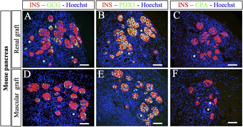
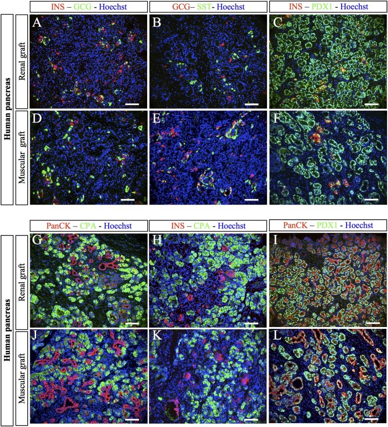
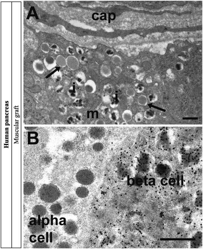
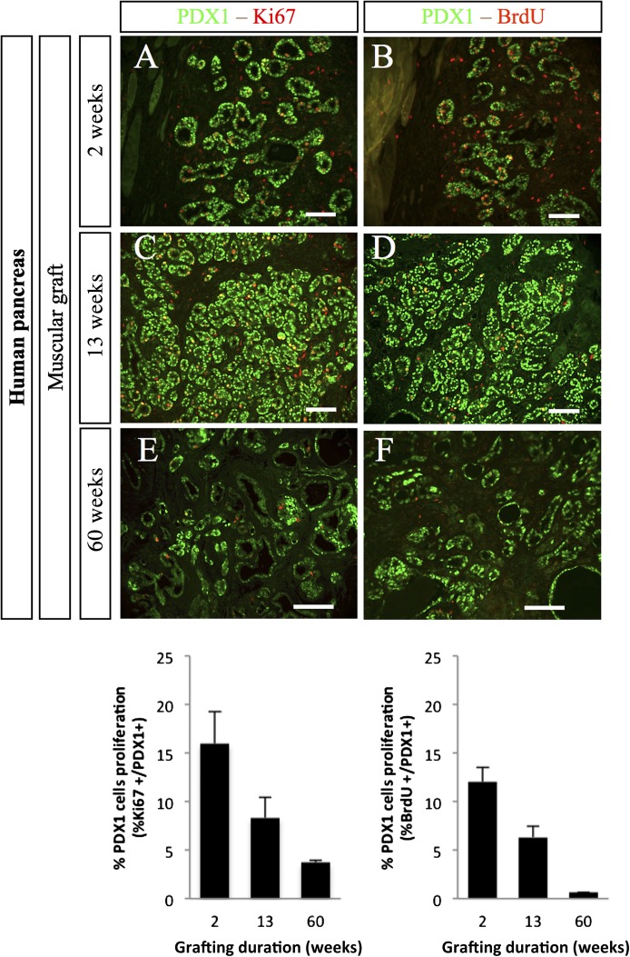
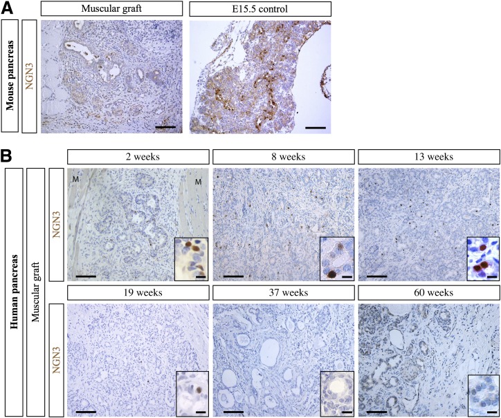
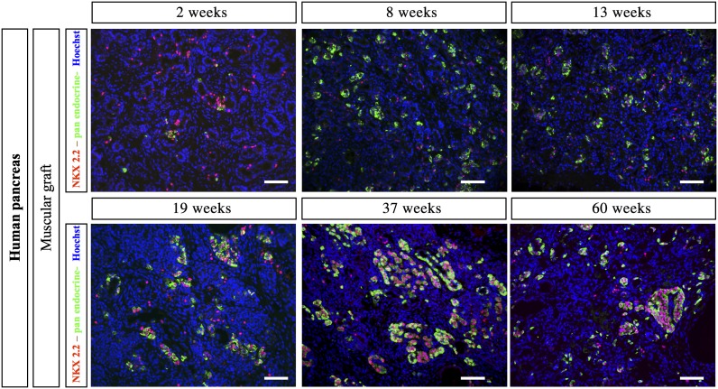
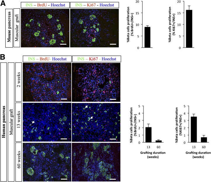
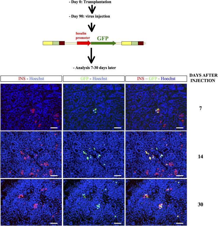
While sporadic human genetic studies have permitted some comparisons between rodent and human pancreatic development, the lack of a robust experimental system has not permitted detailed examination of human pancreatic development. We previously developed a xenograft model of immature human fetal pancreas grafted under the kidney capsule of immune-incompetent mice, which allowed the development of human pancreatic β-cells. Here, we compared the development of human and murine fetal pancreatic grafts either under skeletal muscle epimysium or under the renal capsule. We demonstrated that human pancreatic β-cell development occurs more slowly (weeks) than murine pancreas (days) both by differentiation of pancreatic progenitors and by proliferation of developing β-cells. The superficial location of the skeletal muscle graft and its easier access permitted in vivo lentivirus-mediated gene transfer with a green fluorescent protein-labeled construct under control of the insulin or elastase gene promoter, which targeted β-cells and nonendocrine cells, respectively. This model of engraftment under the skeletal muscle epimysium is a new approach for longitudinal studies, which allows localized manipulation to determine the regulation of human pancreatic development.


Figures

FIG. 1.
FIG. 1.
Development of E12.5 mouse pancreases after grafting. Mouse E12.5 pancreases were grafted either under the kidney capsule (AC) or under the muscle epimysium (DF). Two weeks later, the grafts were removed, sectioned, and stained with anti-insulin, anti-glucagon, anti-PDX1, and anti-CPA antibodies. *Ducts. Scale bars: 100 µm. CPA, carboxypeptidase A; GCG, glucagon; INS, insulin.
FIG. 2.
FIG. 2.
Development of human fetal pancreases after grafting. Human fetal pancreases were grafted either under the kidney capsule (AC and GI) or under the muscle epimysium (DF and JL). Three months later, the grafts were removed, sectioned, and stained with anti-insulin, anti-glucagon, anti-somatostatin, anti-PDX1, anti-CPA, and anti-PanCK antibodies. Scale bars: 100 µm. CPA, carboxypeptidase A; GCG, glucagon; INS, insulin; SST, somatostatin.
FIG. 3.
FIG. 3.
Electron microscopy and gold immunolabeling for insulin in human pancreas 8 months postgrafting in skeletal muscle. A: The graft was well vascularized with capillaries (cap) adjacent to β-cells. β-Cells were well granulated and contained mitochondria (m), mature secretory granules with characteristic crystalline cores of human adult pancreas (i), and some immature granules (arrows). B: Gold immunolabeling for insulin was present over the secretory granules in β-cells but absent from adjacent α-cells. Scale bars: 500 nm.
FIG. 4.
FIG. 4.
Proliferation of PDX1+ cells after grafting of human fetal pancreases under the muscle epimysium. Human fetal pancreases were grafted under the muscle epimysium. At different time points (2, 13, and 60 weeks), mice were injected with BrdU and killed 4 h later. The grafts were removed, sectioned, and stained with anti-PDX1, anti-Ki67, and anti-BrdU antibodies. Top panel: Representative staining at three time points. Scale bars: 100 μm. Lower panel: Quantification of the proportions of PDX1+ cells that were also labeled for either Ki67 or BrdU. n = 4 grafts per group except for at 60 weeks, when n = 2.
FIG. 5.
FIG. 5.
NGN3 expression in mouse and human muscular grafts. A: Mouse E12.5 pancreases were grafted under the muscle epimysium. Two weeks later, the grafts were removed, sectioned, and stained with anti-NGN3 antibodies. Ungrafted E15.5 mouse pancreas was used as a positive control. Scale bars: 100 μm. B: Human fetal pancreases were grafted under the muscle epimysium. At different time points (2, 8, 13, 19, 37, and 60 weeks), the grafts, surrounded by muscle fibers (M), were removed, sectioned, and stained with anti-NGN3 antibodies. Scale bars: 100 μm and 25 μm in the insets.
FIG. 6.
FIG. 6.
NKX2.2 expression in human muscular grafts. Human fetal pancreases were grafted under the muscle epimysium. At different time points (2, 8, 13, 19, 37, and 60 weeks), the grafts were removed, sectioned, and stained with a cocktail of anti-insulin, anti-glucagon, anti-somatostatin, and anti–pancreatic polypeptide antibodies, revealed in green, and anti-NKX2.2 antibodies, revealed in red. Scale bars: 100 μm.
FIG. 7.
FIG. 7.
Proliferation of INS+ cells after grafting of fetal pancreases under the muscle epimysium. Mouse E12.5 (A) and human fetal pancreases (B) were grafted under the muscle epimysium. At different time points, mice were injected with BrdU and killed 4 h later. The grafts were removed, sectioned, and stained with anti-insulin, anti-Ki67, and anti-BrdU antibodies. Representative sections are presented. Arrows point to double-stained cells. Scale bars: 100 μm. Quantification of the proportion of INS+ cells that stained positive for either Ki67 or BrdU is also shown. Quantification was performed on three grafts for mouse pancreas, four grafts for human pancreas at week 13, and two grafts for human pancreas at week 60. INS, insulin.
FIG. 8.
FIG. 8.
Lentivirus-mediated gene transfer into the developing human pancreatic cells for β-cell–specific gene expression. Human fetal pancreases were grafted under the muscle epimysium. Three months later, lentiviral vectors expressing GFP under the control of the insulin promoter were injected into the developing grafts. Grafts were harvested 7, 14, and 30 days postinjection and stained with anti-insulin and anti-GFP antibodies. Scale bars: 100 µm. INS, insulin.

References

    1. Jonsson J, Carlsson L, Edlund T, Edlund H. Insulin-promoter-factor 1 is required for pancreas development in mice. Nature 1994;371:606–609 - PubMed
    1. Stoffers DA, Zinkin NT, Stanojevic V, Clarke WL, Habener JF. Pancreatic agenesis attributable to a single nucleotide deletion in the human IPF1 gene coding sequence. Nat Genet 1997;15:106–110 - PubMed
    1. Ohlsson H, Karlsson K, Edlund T. IPF1, a homeodomain-containing transactivator of the insulin gene. EMBO J 1993;12:4251–4259 - PMC - PubMed
    1. Bhushan A, Itoh N, Kato S, et al. Fgf10 is essential for maintaining the proliferative capacity of epithelial progenitor cells during early pancreatic organogenesis. Development 2001;128:5109–5117 - PubMed
    1. Gradwohl G, Dierich A, LeMeur M, Guillemot F. Neurogenin3 is required for the development of the four endocrine cell lineages of the pancreas. Proc Natl Acad Sci USA 2000;97:1607–1611 - PMC - PubMed

Publication types

MeSH terms

Substances