. 2013 Aug;19(9):1983-9.
doi: 10.1097/MIB.0b013e31829292f7.

Blockade of VEGF receptor-3 aggravates inflammatory bowel disease and lymphatic vessel enlargement

Affiliations

Blockade of VEGF receptor-3 aggravates inflammatory bowel disease and lymphatic vessel enlargement

Giorgia Jurisic et al. Inflamm Bowel Dis. 2013 Aug.

Abstract

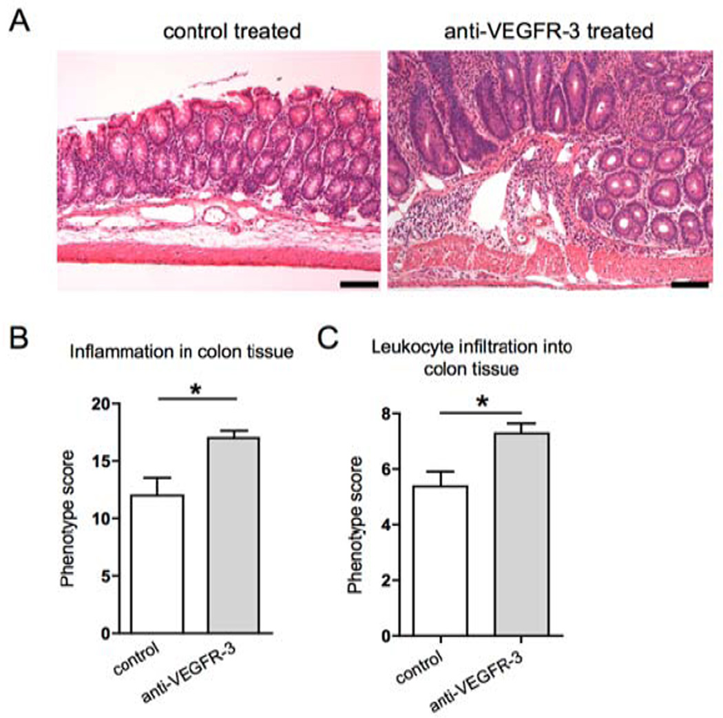
Background: In contrast to the prominent function of the blood vasculature in promoting tissue inflammation, the role of lymphatic vessels in inflammation has been scarcely studied in vivo. To investigate whether modulating lymphatic vessel function might affect the course of chronic inflammation, the major lymphangiogenic receptor, vascular growth factor receptor 3 (VEGFR-3, FLT4), was blocked in an established model of inflammatory bowel disease.

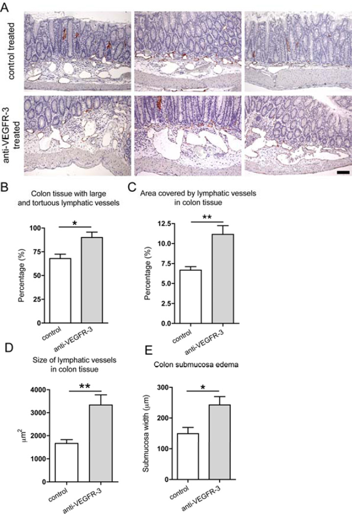
Methods: Interleukin 10 (IL10)-deficient mice that spontaneously develop inflammatory bowel disease were treated with a blocking antibody to VEGFR-3 for 18 days, and the inflammatory changes in colon tissue and the blood and lymphatic vascularization were quantitatively analyzed.

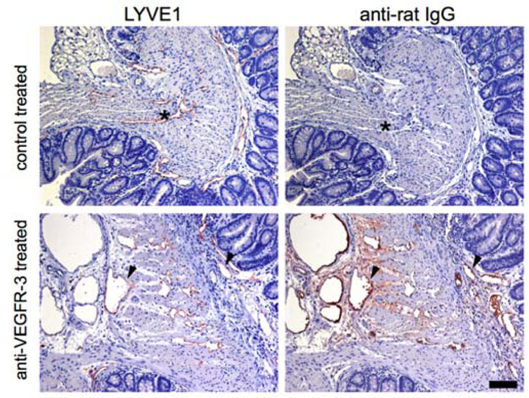
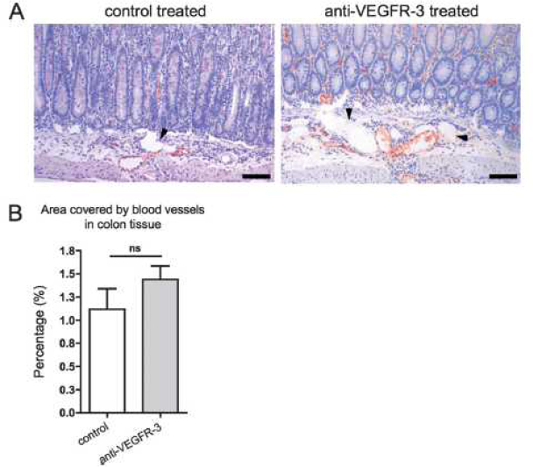
Results: We found a significant increase in the severity of colon inflammation in anti-VEGFR-3-treated mice. This was accompanied by an increased number of enlarged and tortuous lymphatic vessels and edema in colon submucosa, indicating impaired lymphatic function. In contrast, no major effects of the treatment on the blood vasculature were observed.

Conclusions: These results indicate that therapies aimed at promoting lymphatic function, e.g., with prolymphangiogenic factors, such as VEGF-C, might provide a novel strategy for the treatment of inflammatory conditions, such as inflammatory bowel disease.


Figures

Figure 1
Figure 1. Detection of the injected anti-VEGFR-3 blocking antibody bound to lymphatic vessels
Colon sections of mice that received either anti-VEGFR-3 antibody injections or control injections were immunostained with an anti-rat IgG antibody. The rat IgG signal was abundant in the colon lamina propria and submucosa and was co-localized with the LYVE1 signal on lymphatic vessels in serial sections (arrowheads). The rat IgG signal was absent in the colon tissue and lymphatic vessels of control-treated mice (asterisks). Scale bars: 100 µm.
Figure 2
Figure 2. Specific blockade of VEGFR-3 increases colon inflammation and leukocyte infiltration in the Il10−/− mouse model of IBD
(A) Representative images of hematoxylin and eosin stained sections of 8-weeks old C3Bir-Il10−/− mice. Scale bars: 100 µm. (B) Histopathological colitis scoring system: evaluation of inflammation severity, epithelial hyperplasia, mucosal ulceration and extent of area involved along the entire colon. (C) The extent of leukocyte infiltration was evaluated along the entire colon length, inspecting the colon mucosa, lamina propria and submucosa areas (t-test *p ≤ 0.05, n=7–8).
Figure 3
Figure 3. Specific blockade of VEGFR-3 increases lymphatic vessel abnormalities in the Il10−/− mouse model of IBD
(A) Colon sections of 8-weeks old C3Bir-Il10−/− mice were immunostained for LYVE1 (red) and counterstained with hematoxylin. While control mouse colons contained moderately enlarged lymphatic vessels, anti-VEGFR-3 treated mice contained larger areas with dense, enlarged and irregularly shaped lymphatic vessels, surrounded by the edematous tissue. Three representative images are shown, scale bar: 100 µm. Inflammatory changes of lymphatic vessels were evaluated by inspecting the colon tissue for the presence of large and tortuous vessels (B), by determining the area covered by lymphatic vessels in inflamed tissue (C), and by measuring the size of the lymphatic vessels in the inflamed tissue (D). (E) Edema was evaluated by measuring the width of the colon submucosa. (t-test *p ≤ 0.05, **p ≤ 0.01; n=7–8).
Figure 4
Figure 4. Blocking VEGFR-3 does not affect blood vessel density in colon inflammation
(A) Colon sections of 8-weeks old C3Bir-Il10−/− mice were immunostained for the blood vascular marker Meca32 (red) and counterstained with hematoxylin. Arrowheads point to unstained lymphatic vessels. Representative images are shown. Scale bars: 100 µm. (B) Quantification of the tissue area covered by blood vessels revealed no significant difference between control-treated and anti-VEGFR-3-treated mice (t-test p-value: ns=not significant; n=7).

References

    1. Alitalo K. The lymphatic vasculature in disease. Nat Med. 2011;17:1371–1380. - PubMed
    1. Skobe M, Hawighorst T, Jackson DG, et al. Induction of tumor lymphangiogenesis by VEGF-C promotes breast cancer metastasis. Nat Med. 2001;7:192–198. - PubMed
    1. Huggenberger R, Ullmann S, Proulx ST, et al. Stimulation of lymphangiogenesis via VEGFR-3 inhibits chronic skin inflammation. J Exp Med. 2010;207:2255–2269. - PMC - PubMed
    1. Kerjaschki D, Regele HM, Moosberger I, et al. Lymphatic neoangiogenesis in human kidney transplants is associated with immunologically active lymphocytic infiltrates. J Am Soc Nephrol. 2004;15:603–612. - PubMed
    1. Kaipainen A, Korhonen J, Mustonen T, et al. Expression of the fms-like tyrosine kinase 4 gene becomes restricted to lymphatic endothelium during development. Proc Natl Acad Sci U S A. 1995;92:3566–3570. - PMC - PubMed

Publication types

MeSH terms

Substances