Review
. 2013 Aug;9(8):429-44.
doi: 10.1038/nrneurol.2013.126. Epub 2013 Jul 9.

The clinical maze of mitochondrial neurology

Affiliations
Review

The clinical maze of mitochondrial neurology

Salvatore DiMauro et al. Nat Rev Neurol. 2013 Aug.

Abstract

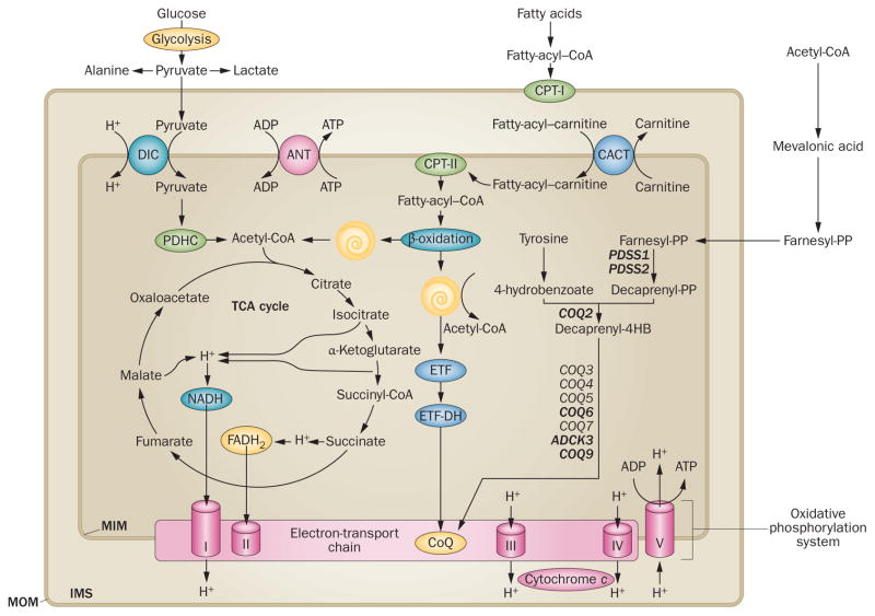
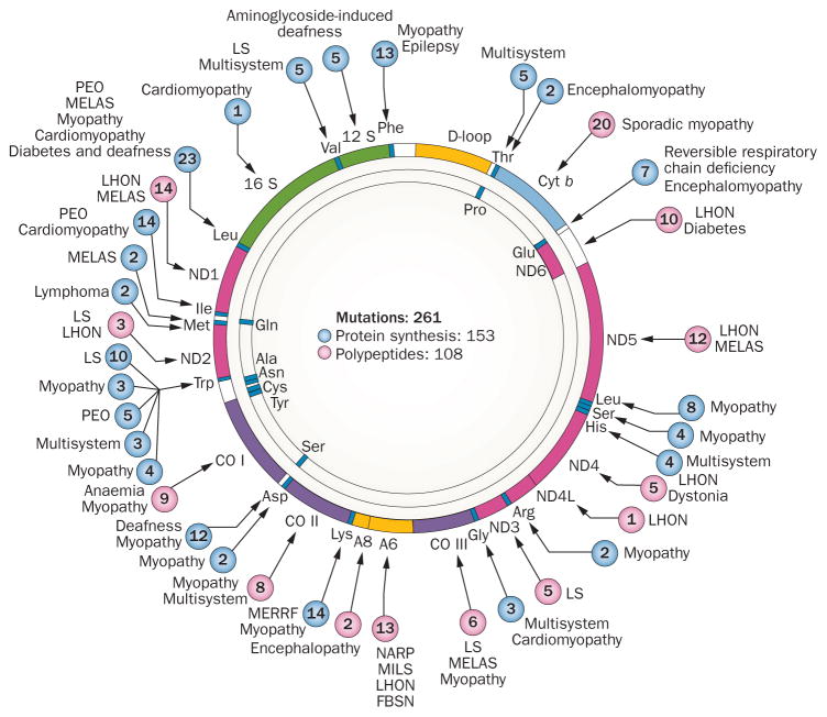
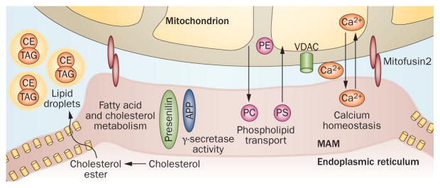
Mitochondrial diseases involve the respiratory chain, which is under the dual control of nuclear and mitochondrial DNA (mtDNA). The complexity of mitochondrial genetics provides one explanation for the clinical heterogeneity of mitochondrial diseases, but our understanding of disease pathogenesis remains limited. Classification of Mendelian mitochondrial encephalomyopathies has been laborious, but whole-exome sequencing studies have revealed unexpected molecular aetiologies for both typical and atypical mitochondrial disease phenotypes. Mendelian mitochondrial defects can affect five components of mitochondrial biology: subunits of respiratory chain complexes (direct hits); mitochondrial assembly proteins; mtDNA translation; phospholipid composition of the inner mitochondrial membrane; or mitochondrial dynamics. A sixth category-defects of mtDNA maintenance-combines features of Mendelian and mitochondrial genetics. Genetic defects in mitochondrial dynamics are especially important in neurology as they cause optic atrophy, hereditary spastic paraplegia, and Charcot-Marie-Tooth disease. Therapy is inadequate and mostly palliative, but promising new avenues are being identified. Here, we review current knowledge on the genetics and pathogenesis of the six categories of mitochondrial disorders outlined above, focusing on their salient clinical manifestations and highlighting novel clinical entities. An outline of diagnostic clues for the various forms of mitochondrial disease, as well as potential therapeutic strategies, is also discussed.


Conflict of interest statement

Competing interests

The authors declare no competing interests.

Figures

Figure 1
Figure 1
The mitochondrial respiratory chain. Electrons derived from cellular dehydrogenases in the Krebs cycle and in β-oxidation spirals are passed ‘horizontally’ along four protein complexes and two small carriers (the electron transport chain) that are embedded in the MIM. The electrons travel from complex I (an NADH dehydrogenase) and complex II (a succinate dehydrogenase) to CoQ10 (a small mobile electron carrier also known as ubiquinone), then to complex III (ubiquinone oxidoreductase), cytochrome c (another small mobile electron carrier) and complex IV (cytochrome c oxidase), ultimately producing water. Concomitant with this horizontal flow of electrons, ‘vertical’ vectorial transport of dehydrogenase-derived protons from the matrix across the MIM into the intermembrane space takes place. This process creates an electrochemical proton gradient across the MIM that is used to drive complex V (F0F1-ATP synthase), a rotary motor that converts ADP to ATP. Conventionally, the five complexes comprise the oxidative phosphorylation system. The biosynthetic pathway of CoQ10, beginning with acetyl-CoA, is shown on the right; mutated biosynthetic genes that cause deficiency are show in bold text. Abbreviations: ANT, adenine nucleotide translocator; CACT, carnitine–primary CoQ10 acylcarnitine translocator; CoA, coenzyme A; CoQ10, coenzyme Q10; CPT, carnitine palmitoyltransferase; DIC, dicarboxylate carrier; ETF, electron-transfer flavoprotein; ETF-DH, ETF-dehydrogenase; FAD, flavin adenine dinucleotide; IMS, intermembrane space; MIM, mitochondrial inner membrane; MOM, mitochondrial outer membrane; PDHC, pyruvate dehydrogenase complex; TCA, tricarboxylic acid. Image first printed in Molecular Neurology.
Figure 2
Figure 2
The mitochondrial morbidity map. Schematic map of the 16,569-bp mtDNA, in which coloured sections represent protein-coding genes: seven subunits of complex I (ND; pink sections); one subunit of complex III (cyt b; light blue section); three subunits of cytochrome c oxidase (CO; purple sections); two subunits of ATP synthase (A6 and A8; yellow sections): 12 S and 16 S ribosomal RNA (green sections); and 22 transfer RNAs identified by three-letter codes for the corresponding amino acids (blue sections). Diseases due to mutations in genes that impair protein synthesis are indicated as blue circles. Mutated genes that encode respiratory chain proteins are indicated as pink circles. Numbers in circles represent number of mutations reported at the given site. Abbreviations: Cyt b, cytochrome b; FBSN, familial bilateral striatal necrosis; LHON, Leber hereditary optic neuropathy; LS, Leigh syndrome; MELAS, mitochondrial encephalomyopathy, lactic acidosis, and stroke-like episodes; MERRF, myoclonus epilepsy with ragged-red fibres; MILS, maternally inherited Leigh syndrome; NARP, neuropathy, ataxia and retinitis pigmentosa; ND, NADH-dehydrogenase (complex I); PEO, progressive external ophthalmoplegia.
Figure 3
Figure 3
Structure and function of the MAM. The ER communicates with mitochondria via the MAM, a specialized subregion of the ER with the characteristics of a lipid raft. The MAM is the regulatory site in the cell for phospholipid metabolism, cholesterol ester and fatty acid metabolism, lipid droplet formation, calcium homeostasis, and mitochondrial dynamics. Abbreviations: APP, amyloid precursor protein; CE, cholesteryl ester; ER, endoplasmic reticulum; MAM, mitochondria-associated ER membrane; PC, phosphatidylcholine; PE, phosphatidylethanolamine; PS, phosphatidylserine; TAG, triacylglycerol, VDAC, voltage-dependent anion-selective channel. Original image courtesy of E. Area-Gomez, Columbia University, NY, USA.
Figure 4
Figure 4
Muscle biopsy in patients with mitochondrial disease. a | In a patient with MERRF (harbouring a mitochondrial DNA point mutation that impairs mitochondrial protein synthesis), two ragged-red fibres are evident with the modified Gomori trichrome stain. bd | Using the SDH stain, the same fibres appear ‘ragged blue’ (part b) but are COX-negative (part c), and stain blue with the combined COX and SDH stain (COX-deficient fibres stain a lighter blue; part d). Abbreviations: MERRF, myoclonus epilepsy with ragged-red fibres; SDH, succinate dehydrogenase. Image first printed in Molecular Neurology.

Similar articles

Cited by

References

    1. Luft R, Ikkos D, Palmieri G, Ernster L, Afzelius B. A case of severe hypermetabolism of nonthyroid origin with a defect in the maintenance of mitochondrial respiratory control: a correlated clinical, biochemical, and morphological study. J Clin Invest. 1962;41:1776–1804. - PMC - PubMed
    1. Shapira Y, Harel S, Russell A. Mitochondrial encephalomyopathies: a group of neuromuscular disorders with defects in oxidative metabolism. Isr J Med Sci. 1977;13:161–164. - PubMed
    1. Holt IJ, Harding AE, Morgan-Hughes JA. Deletions of muscle mitochondrial DNA in patients with mitochondrial myopathies. Nature. 1988;331:717–719. - PubMed
    1. Wallace DC, et al. Mitochondrial DNA mutation associated with Leber’s hereditary optic neuropathy. Science. 1988;242:1427–1430. - PubMed
    1. Schon EA, DiMauro S, Hirano M. Human mitochondrial DNA: roles of inherited and somatic mutations. Nat Rev Genet. 2012;13:878–890. - PMC - PubMed

Publication types

MeSH terms