. 2013 Jul 24;19(1):212-22.
doi: 10.2119/molmed.2013.00036.

Corticotropin-releasing factor receptor 2 mediates sex-specific cellular stress responses

Affiliations

Corticotropin-releasing factor receptor 2 mediates sex-specific cellular stress responses

Eric Kubat et al. Mol Med. .

Abstract

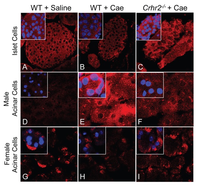
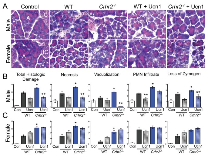
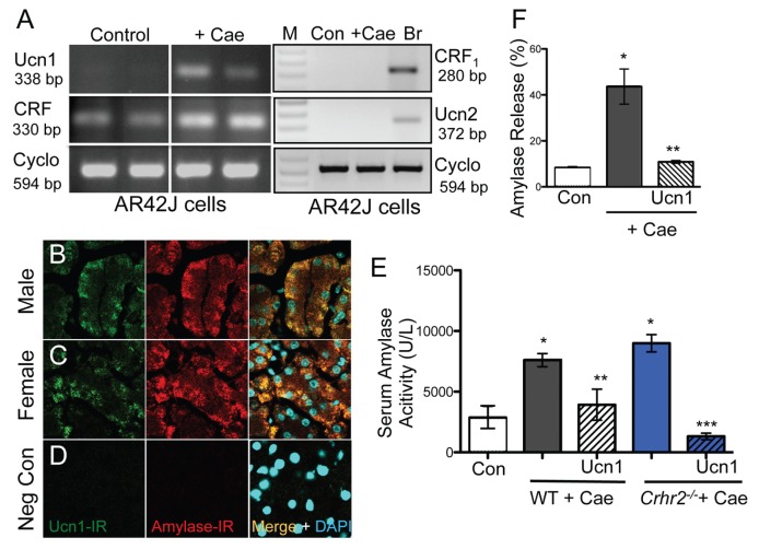
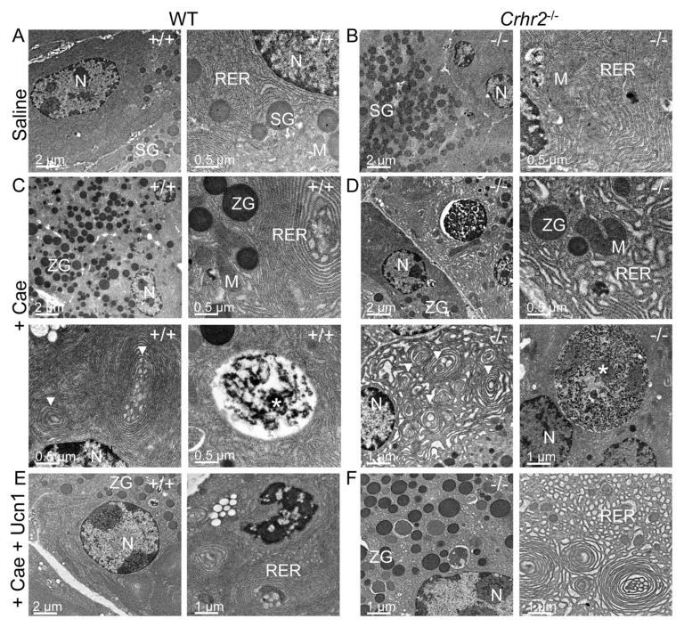
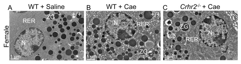
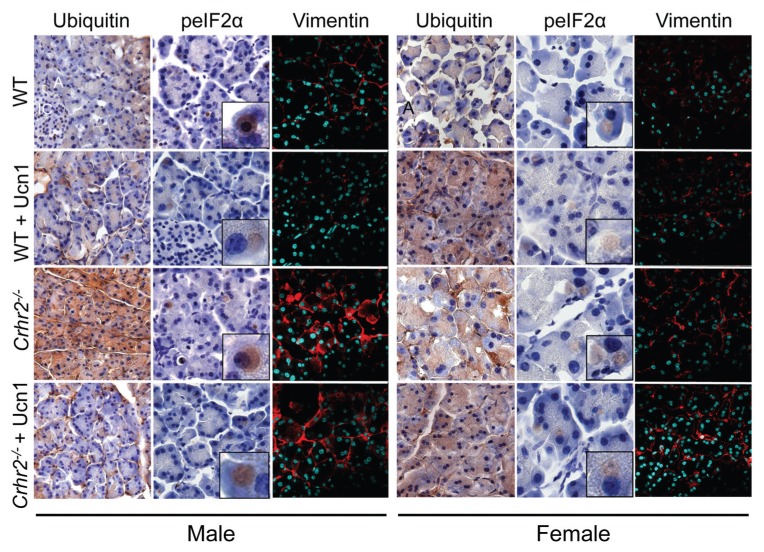
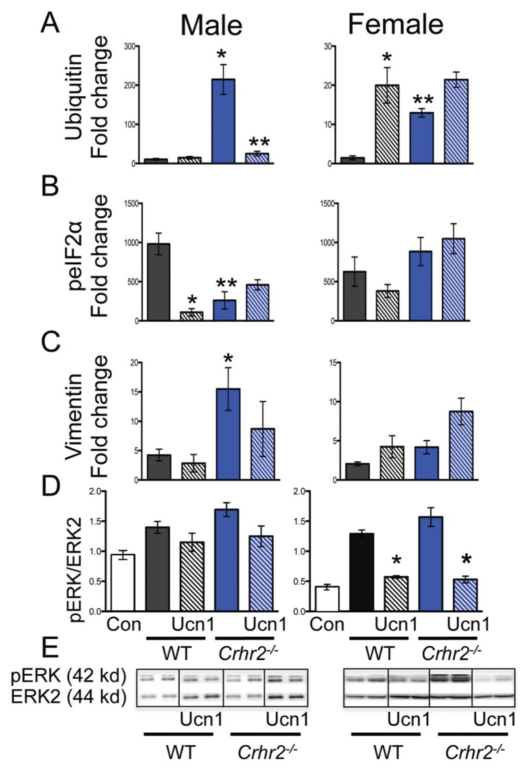
Although females suffer twice as much as males from stress-related disorders, sex-specific participating and pathogenic cellular stress mechanisms remain uncharacterized. Using corticotropin-releasing factor receptor 2-deficient (Crhr2-/-) and wild-type (WT) mice, we show that CRF receptor type 2 (CRF2) and its high-affinity ligand, urocortin 1 (Ucn1), are key mediators of the endoplasmic reticulum (ER) stress response in a murine model of acute pancreatic inflammation. Ucn1 was expressed de novo in acinar cells of male, but not female WT mice during acute inflammation. Upon insult, acinar Ucn1 induction was markedly attenuated in male but not female Crhr2-/- mice. Crhr2-/- mice of both sexes show exacerbated acinar cell inflammation and necrosis. Electron microscopy showed mild ER damage in WT male mice and markedly distorted ER structure in Crhr2-/- male mice during pancreatitis. WT and Crhr2-/- female mice showed similarly distorted ER ultrastructure that was less severe than distortion seen in Crhr2-/- male mice. Damage in ER structure was accompanied by increased ubiquitination, peIF2, and mistargeted localization of vimentin in WT mice that was further exacerbated in Crhr2-/- mice of both sexes during pancreatitis. Exogenous Ucn1 rescued many aspects of histological damage and cellular stress response, including restoration of ER structure in male WT and Crhr2-/- mice, but not in females. Instead, females often showed increased damage. Thus, specific cellular pathways involved in coping and resolution seem to be distinct to each sex. Our results demonstrate the importance of identifying sex-specific pathogenic mechanisms and their value in designing effective therapeutics.


Figures

Figure 1
Figure 1
De novo induction of Ucn1 in exocrine acinar cells. Pancreatic sections were stained with anti-Ucn1 antibody and visualized under a confocal microscope. Representative sections are shown from saline-treated WT (A, D, G), caerulein-treated (+ Cae) WT (B, E, H), and caerulein-treated Crhr2/ (C, F, I) pancreatic sections (60×; inset magnification: 173×) from male and female mice (n = 6/group).
Figure 2
Figure 2
CRF2 actions are key determinants of an inflammatory insult. Representative light micrographs (40×; A, acinar cells, IC, islet cells) of H&E stained pancreas sections from male and female saline-treated WT, caerulein-treated WT, and caerulein-treated Crhr2/mice (A). Graphical representation of histologic damage scores graded by a pathologist is shown. Duplicate sections were scored for necrosis, vacuolization, polymorphonuclear cell infiltration and zymogen granule formation in vehicle-treated WT (Con, white bars), caerulein-treated WT (grey bars) and caerulein-treated Crhr2/ (blue bars) mice. Severity scores increased in all histopathologic categories for male Crhr2/ mice (*p < 0.05 versus WT) (B). Total score, necrosis and vacuolization increased in female Crhr2/ mice (*p < 0.05) (C). Ucn1 treatment (hashed grey and blue bars) significantly decreased all scores for male Crhr2−/− except PMN infiltrate, which was decreased in male WT mice (**p < 0.05). Ucn1 treatment either worsened histologic damage in female WT and Crhr2/ mice or had no effect on variables examined (C). Statistical analyses were performed between same-sex groups by using ANOVA, followed by Bonferroni posttests. Data represent score ± SEM of 12 mice/group.
Figure 3
Figure 3
Ucn1 colocalizes with amylase in acinar cells. Ucn1 mRNA was induced de novo in acinar AR42J cells treated with 10−7 mol/L caerulein for 1 h. CRF mRNA expression was present under basal conditions and was not induced de novo. Ucn2 and CRF1 mRNA were not detected at baseline (Con) or after caerulein treatment in acinar cells, but were present in brain (Br) mRNA. Cyclophilin was used as a normalization control (A). Caerulein treatment increased amylase-IR in acinar cells in WT mice and colocalized with Ucn1-IR in male and female mice (B, C), respectively (63×); sections stained without any primary antibody served as negative controls (D). Serum amylase activity increased over basal (control) levels (*p < 0.05) in WT (grey bars) and Crhr2/ (blue bars) mice (E), and Ucn1 treatment (hashed grey and blue bars) significantly decreased amylase release (**p < 0.05 versus WT; ***p < 0.05 versus Crhr2/). Amylase release from AR42J cells (F). Caerulein treatment increased amylase release (*p < 0.05) and 10−7 mol/L Ucn1 treatment decreased release (**p < 0.05). Statistical analyses were by ANOVA. Data represent mean ± SEM of three replicates per group.
Figure 4
Figure 4
CRF2 is central to the maintenance of organelle integrity and the endoplasmic reticulum stress responses. Representative electron microscopy (EM) shows normal acinar cell ultrastructure in saline-treated WT (A) or saline-treated Crhr2/ (B) mice (n = 4/group) with normal appearing acinar cell ultrastructure. During pancreatitis, WT male mice show a marked increase in zymogen granule (ZG) formation, mild expansion of rough ER and intraluminal material (C), consistent with ER stress. Infrequent, small autophagic bodies (*) containing densely staining material were found. During pancreatitis, Crhr2−/− mice show markedly dilated rough ER (D); ER whorls and increased intraluminal inclusion bodies were visible, all of which were increased relative to what was seen in WT pancreatitis. Frequent, multiple large autophagic bodies (white *) that contained numerous ribosomes and membrane components were seen. (M, mitochondria; N, nucleus; RER, rough endoplasmic reticulum; SG, secretory granules; ZG, zymogen granule). Exogenous Ucn1 treatment during pancreatitis markedly reduces ER ultrastructure damage and number of autophagic bodies in both WT and Crhr2−/− male mice (E, F).
Figure 5
Figure 5
Lack of Crhr2−/− in female mice results in a milder ER phenotype. WT female controls show normal organelle ultrastructure (A) similar to their male littermates. When pancreatitis is induced, females, unlike their male counterparts, show milder ER ultrastructure damage in both WT (B) and Crhr2/ (C) genotypes. (N: nucleus, RER: rough endoplasmic reticulum, ZG: zymogen granule).
Figure 6
Figure 6
CRF2 disrupts the ER stress response. Pancreatic sections immunostained for ER stress pathway proteins. Representative light micrographs (60×; inset: 300×; A, acinar cells, IC, islet cells) immunostained for ubiquitin and peIF2α in WT and Crhr2/ male and female control mice or mice treated with caerulein alone or caerulein + Ucn1 are shown. Mistargeting of vimentin is evident in Crhr2/ mice of both sexes (magnification: 63×; nuclei stained with 4′,6-diamidino-2-phenylindole (DAPI); n = 6/group).
Figure 7
Figure 7
ER stress response is restored after Ucn1 treatment. Graphical representation of fold-change in staining intensity for ubiquitin (A), peIF2α (B), and vimentin (C) over saline-treated controls is shown (*p < 0.05, WT versus Crhr2/). Simultaneous Ucn1 treatment decreased ubiquitin levels in male Crhr2−/− mice (** p < 0.05) and peIF2α levels in male WT mice (*p < 0.05) (A, B). Western blot analysis of pancreatic lysates was performed (D, E), and representative Western blot from two mice per group is shown (E). Quantitation of pERK1/2 normalized to total ERK showed significant increases in pERK1/2 in male and female WT and Crhr2/ mice (D, *p < 0.05). Ucn1 treatment did not affect pERK levels in male WT or Crhr2/ mice, but significantly (**p < 0.05) reduced pERK levels in female WT and Crhr2/ mice. Statistical analyses were performed between same-sex groups by using ANOVA. Data represent mean ± SEM of 12 mice per group.

Similar articles

Cited by

References

    1. Muglia L, Jacobson L, Dikkes P, Majzoub JA. Corticotropin-releasing hormone deficiency reveals major fetal but not adult glucocorticoid need. Nature. 1995;373:427–32. - PubMed
    1. Slominski A, et al. Cutaneous expression of corticotropin-releasing hormone (CRH), urocortin, and CRH receptors. FASEB J. 2001;15:1678–93. - PubMed
    1. Chen A, Blount A, Vaughan J, Brar B, Vale W. Urocortin II gene is highly expressed in mouse skin and skeletal muscle tissues: localization, basal expression in corticotropin-releasing factor receptor (CRFR) 1- and CRFR2-null mice, and regulation by glucocorticoids. Endocrinology. 2004;145:2445–57. - PubMed
    1. Baigent SM. Peripheral corticotropin-releasing hormone and urocortin in the control of the immune response. Peptides. 2001;22:809–20. - PubMed
    1. Wu Y, Xu Y, Zhou H, Tao J, Li S. Expression of urocortin in rat lung and its effect on pulmonary vascular permeability. J Endocrinol. 2006;189:167–78. - PubMed

Publication types