. 2013 Sep;126(3):385-99.
doi: 10.1007/s00401-013-1149-y. Epub 2013 Jul 9.

Modeling key pathological features of frontotemporal dementia with C9ORF72 repeat expansion in iPSC-derived human neurons

Affiliations

Modeling key pathological features of frontotemporal dementia with C9ORF72 repeat expansion in iPSC-derived human neurons

Sandra Almeida et al. Acta Neuropathol. 2013 Sep.

Erratum in

  • Acta Neuropathol. 2014 Jun;127(6):941

Abstract

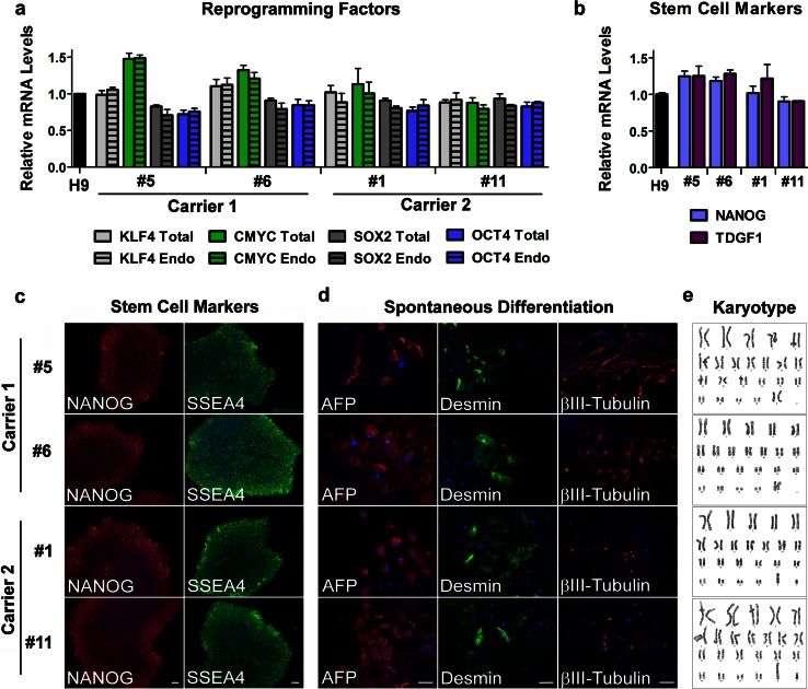
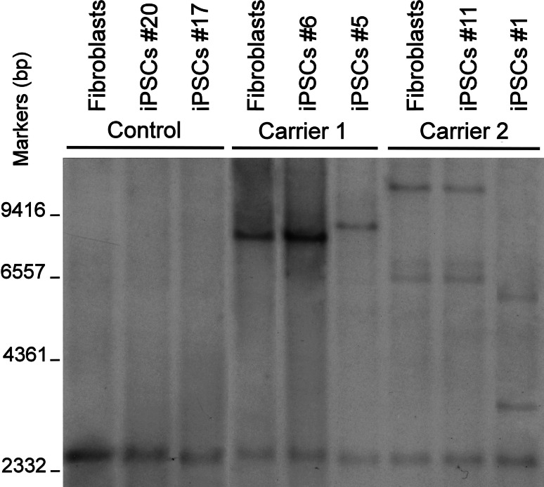
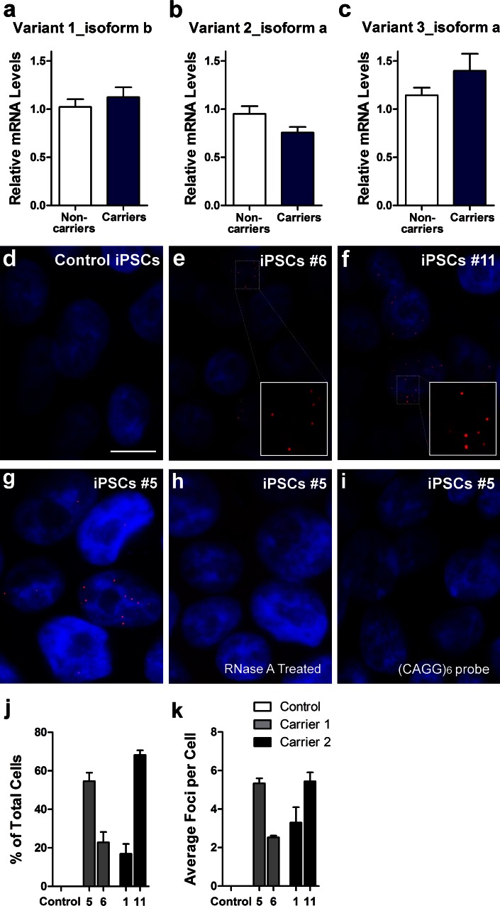
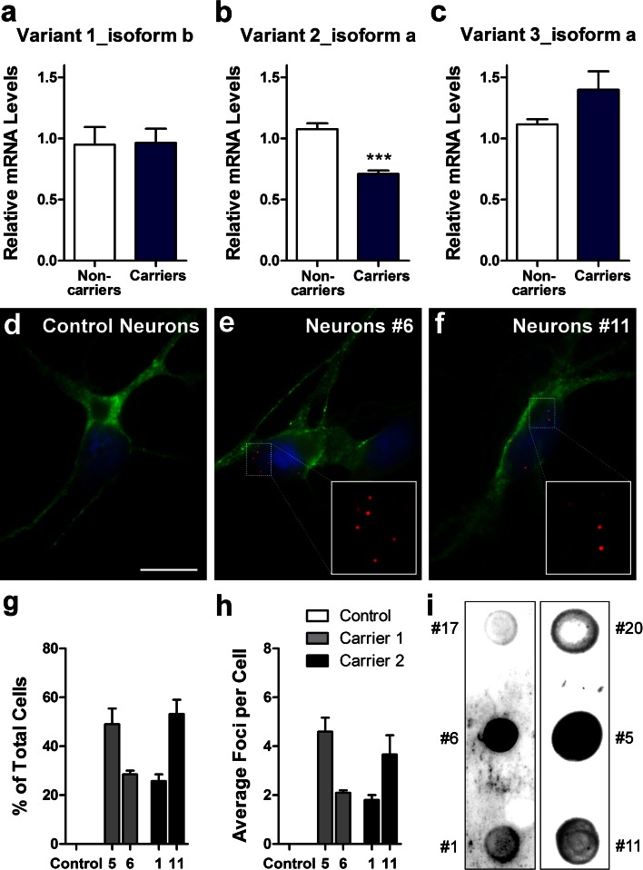
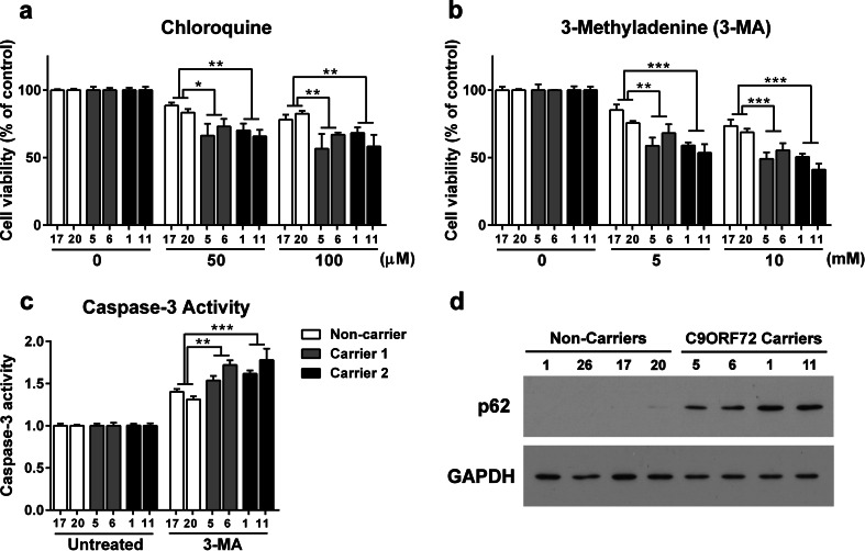
The recently identified GGGGCC repeat expansion in the noncoding region of C9ORF72 is the most common pathogenic mutation in patients with frontotemporal dementia (FTD) or amyotrophic lateral sclerosis (ALS). We generated a human neuronal model and investigated the pathological phenotypes of human neurons containing GGGGCC repeat expansions. Skin biopsies were obtained from two subjects who had >1,000 GGGGCC repeats in C9ORF72 and their respective fibroblasts were used to generate multiple induced pluripotent stem cell (iPSC) lines. After extensive characterization, two iPSC lines from each subject were selected, differentiated into postmitotic neurons, and compared with control neurons to identify disease-relevant phenotypes. Expanded GGGGCC repeats exhibit instability during reprogramming and neuronal differentiation of iPSCs. RNA foci containing GGGGCC repeats were present in some iPSCs, iPSC-derived human neurons and primary fibroblasts. The percentage of cells with foci and the number of foci per cell appeared to be determined not simply by repeat length but also by other factors. These RNA foci do not seem to sequester several major RNA-binding proteins. Moreover, repeat-associated non-ATG (RAN) translation products were detected in human neurons with GGGGCC repeat expansions and these neurons showed significantly elevated p62 levels and increased sensitivity to cellular stress induced by autophagy inhibitors. Our findings demonstrate that key neuropathological features of FTD/ALS with GGGGCC repeat expansions can be recapitulated in iPSC-derived human neurons and also suggest that compromised autophagy function may represent a novel underlying pathogenic mechanism.


Figures

Fig. 1
Fig. 1
Generation and characterization of iPSC lines from carriers of C9ORF72 expanded repeats. Total and endogenous (Endo) mRNA levels of the reprogramming factors KLF4, CMYC, SOX2, and OCT4 in iPSC lines from carriers 1 and 2 relative to the values in human embryonic stem cell line H9 were assessed by qRT-PCR. Values are mean ± SEM (a). The expression of the pluripotency markers NANOG and TDGF1 (or CRIPTO) was measured at the mRNA level. The values from H9 cells were set to 1. Values are mean ± SEM (b). Immunofluorescence analysis of pluripotency markers NANOG and SSEA4 in iPSC lines from carriers 1 and 2 is shown and cell nuclei were counterstained with Hoechst (blue). Scale bar 50 μm (c). After in vitro spontaneous differentiation of iPSC lines into cells of three embryonic germ layers, cells were immunostained with α-fetoprotein (AFP, endoderm), desmin (mesoderm), βIII-tubulin (ectoderm), and Hoechst (nuclei). Scale bar 50 μm (d). All iPSC lines maintained a normal karyotype (e)
Fig. 2
Fig. 2
Southern blot analysis of C9ORF72 alleles in fibroblasts and two iPSC lines (passage 24) derived from each individual. One representative blot is shown here
Fig. 3
Fig. 3
GGGGCC repeats do not affect neuronal differentiation of iPSCs. MAP-2-positive neurons with or without GGGGCC repeat expansions after neuronal differentiation, using methods described in Almeida et al. [3]. Nuclear staining is shown in blue. Scale bar 50 μm (a). The percentage of MAP-2-positive neurons (b) and GFAP-positive astrocytes (c) in 2 week-old cultures are shown. Cells positive for VGLUT1 (d), GABA (e), and TH (f) (glutamatergic, GABAergic and dopaminergic markers, respectively) were counted as a percentage of MAP-2+ cells. On average, 200 cells were analyzed per experiment (n = 3 independent cultures). Values are mean ± SEM. Representative depolarization-evoked action potentials from neurons at baseline and after 2 min application of tetrodotoxin (0.5 μM) are shown. Action potentials were elicited by current injections from +400 to 0 pA in 100 pA steps (n = 10–14 for each line) (g). Representative spontaneous EPSCs at baseline and after 2 min application of CNQX (10 μM). Neurons were held at −60 mV under voltage-clamp (n = 10–11 from each line) (h)
Fig. 4
Fig. 4
GGGGCC repeats instability during neuronal differentiation of iPSCs. Southern blot analysis of iPSC line 20 of control (a), line 6 of carrier 1 (b), and line 11 of carrier 2 (c) at different passages and their differentiated neurons. The asterisks indicate the mutant C9ORF72 alleles in neurons. Brains from a control subject and two C9ORF72 repeat expansion carriers show repeats of different lengths (d)
Fig. 5
Fig. 5
C9ORF72 repeat expansions form RNA foci in iPSCs. Expression levels of C9ORF72 variant 1 (NM_145005.5, isoform b) (a), variant 2 (NM_018325.3, isoform a) (b) and variant 3 (NM_001256054.1, isoform a) (c) in iPSC lines from two non-carriers and two expanded repeat carriers were assessed by qRT-PCR. Fluorescence in situ hybridization (FISH) analysis was done on control iPSC line 20 (d), carrier 1 line 6 iPSCs (e), carrier 2 line 11 iPSCs (f) using a cy3-conjugated (GGCCCC)4 probe. RNA foci (red) were found in the nucleus (blue) of carriers 1 and 2 but not in control cells. Treatment of iPSCs with RNase A after fixation leads to loss of foci (h), indicating that the foci are indeed made of RNA. Representative images of iPSCs from carrier 1 (line 5) that were left untreated (g) or treated with RNase A (h) for 20 min at room temperature. Red RNA foci containing GGGGCC repeats; blue nuclei (DAPI). Cells did not show foci when a Cy3-conjugated (CAGG)6 probe was used as the negative control probe (i). Scale bar 10 μm. Quantifications of the percentage of iPSCs displaying foci (j) and the average number of foci per cell (k) are shown as mean ± SEM
Fig. 6
Fig. 6
C9ORF72 repeat expansions form RNA foci in patient iPSCs-derived neurons. Expression levels of C9ORF72 variant 1 (NM_145005.5, isoform b) (a), variant 2 (NM_018325.3, isoform a) (b) and variant 3 (NM_001256054.1, isoform a) (c) in iPSC-derived neurons from two non-carriers and two expanded repeat carriers were assessed by qRT-PCR. Values are mean ± SEM, *** p < 0.001 (Student’s t test). FISH analysis was done on control iPSC-derived neurons (d), carrier 1 line 6 iPSC-derived neurons (e), carrier 2 line 11 iPSC-derived neurons (f) using a cy3-conjugated (GGCCCC)4 probe. Green MAP2. Blue DAPI. Scale bar 10 μm. Quantifications of the percentage of neurons displaying foci (g) and the average number of foci per cell (h) are presented as mean ± SEM, based on analysis of neurons derived from three independent differentiation experiments. Gly-Pro dipeptide repeats are detected by dot blot analysis in neurons of carrier 1 (iPSC lines 5 and 6) and carrier 2 (iPSC lines 1 and 11) (i)
Fig. 7
Fig. 7
GGGGCC repeat expansions exacerbate susceptibility to inhibition of the autophagy pathway. Cell viability of human neurons after exposure to chloroquine (a) and 3-methyladenine (3-MA) (b) for 24 h. Values are expressed as a percentage of the untreated cells (control) (n = 3 independent cultures) (a, b). Caspase-3-like activity after exposure to 5 mM 3-MA for 24 h (c). In all panels, values are mean ± SEM. *p < 0.05, **p < 0.01, ***p < 0.001 (One-way ANOVA). Representative Western blot showing p62 protein levels in neurons of two non-carriers (PGRN S116X: lines 1 and 26, and control: lines 17 and 20) and two repeat expansions carriers (carrier 1: lines 5 and 6, and carrier 2: lines 1 and 11) (d)

Similar articles

Cited by

References

    1. Abdelmohsen K, Gorospe M. RNA-binding protein nucleolin in disease. RNA Biol. 2012;9:799–808. doi: 10.4161/rna.19718. - DOI - PMC - PubMed
    1. Abyzov A, Mariani J, Palejev D, et al. Somatic copy number mosaicism in human skin revealed by induced pluripotent stem cells. Nature. 2012;492:438–442. doi: 10.1038/nature11629. - DOI - PMC - PubMed
    1. Almeida S, Zhang Z, Copplola G, et al. Induced pluripotent stem cell models of progranulin-deficient frontotemporal dementia uncover specific reversible neuronal defects. Cell Rep. 2012;2:789–798. doi: 10.1016/j.celrep.2012.09.007. - DOI - PMC - PubMed
    1. Al-Sarraj S, King A, Troakes C, et al. p62 positive, TDP-43 negative, neuronal cytoplasmic and intranuclear inclusions in the cerebellum and hippocampus define the pathology of C9orf72-linked FTLD and MND/ALS. Acta Neuropathol. 2011;122:691–702. doi: 10.1007/s00401-011-0911-2. - DOI - PubMed
    1. Amaravadi RK, Yu D, Lum JJ, et al. Autophagy inhibition enhances therapy-induced apoptosis in a Myc-induced model of lymphoma. J Clin Invest. 2007;117:326–336. doi: 10.1172/JCI28833. - DOI - PMC - PubMed

Publication types

MeSH terms

Supplementary concepts