. 2013 Dec;74(6):805-14.
doi: 10.1002/ana.23971.

Mutations in γ adducin are associated with inherited cerebral palsy

Affiliations

Mutations in γ adducin are associated with inherited cerebral palsy

Michael C Kruer et al. Ann Neurol. 2013 Dec.

Abstract

Objective: Cerebral palsy is estimated to affect nearly 1 in 500 children, and although prenatal and perinatal contributors have been well characterized, at least 20% of cases are believed to be inherited. Previous studies have identified mutations in the actin-capping protein KANK1 and the adaptor protein-4 complex in forms of inherited cerebral palsy, suggesting a role for components of the dynamic cytoskeleton in the genesis of the disease.

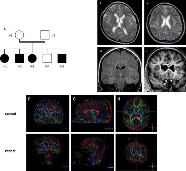
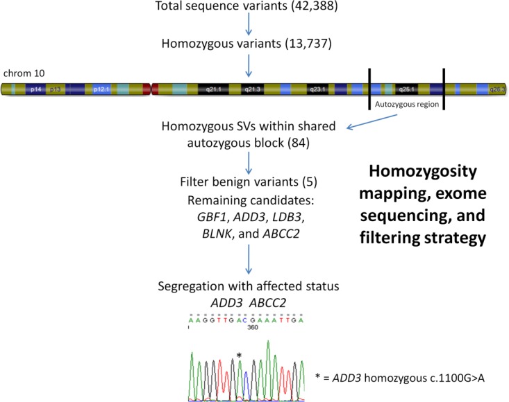
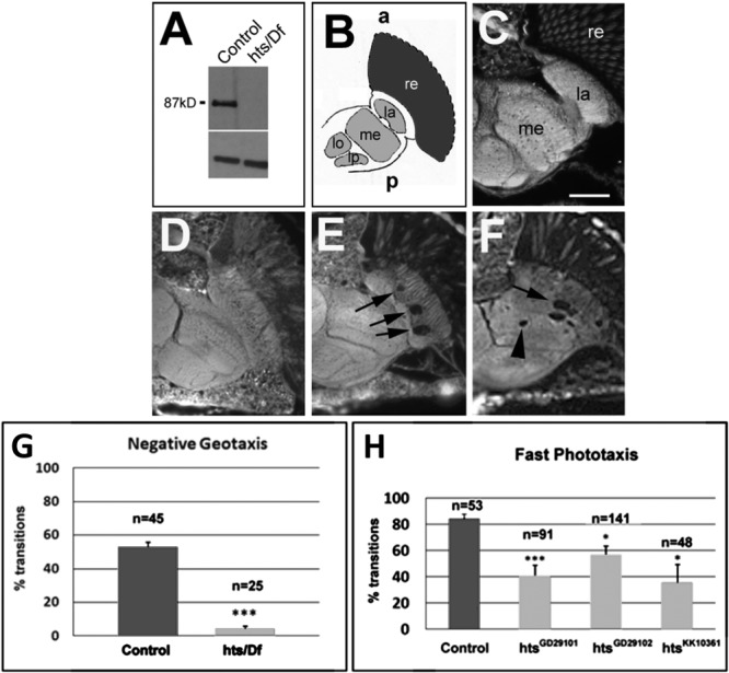
Methods: We studied a multiplex consanguineous Jordanian family by homozygosity mapping and exome sequencing, then used patient-derived fibroblasts to examine functional consequences of the mutation we identified in vitro. We subsequently studied the effects of adducin loss of function in Drosophila.

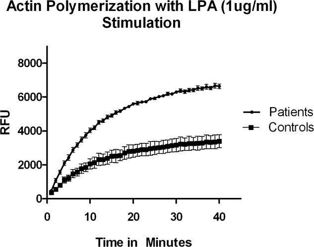
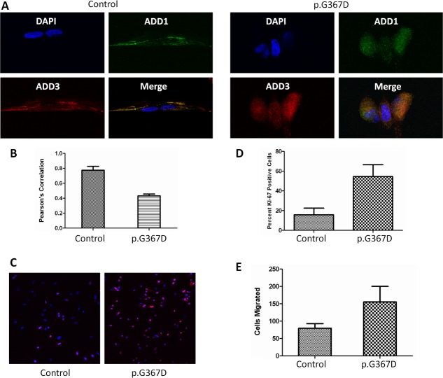
Results: We identified a homozygous c.1100G>A (p.G367D) mutation in ADD3, encoding gamma adducin in all affected members of the index family. Follow-up experiments in patient fibroblasts found that the p.G367D mutation, which occurs within the putative oligomerization critical region, impairs the ability of gamma adducin to associate with the alpha subunit. This mutation impairs the normal actin-capping function of adducin, leading to both abnormal proliferation and migration in cultured patient fibroblasts. Loss of function studies of the Drosophila adducin ortholog hts confirmed a critical role for adducin in locomotion.

Interpretation: Although likely a rare cause of cerebral palsy, our findings indicate a critical role for adducins in regulating the activity of the actin cytoskeleton, suggesting that impaired adducin function may lead to neuromotor impairment and further implicating abnormalities of the dynamic cytoskeleton as a pathogenic mechanism contributing to cerebral palsy.


Figures

FIGURE 1
FIGURE 1
Pedigree and neuroimaging findings of index family. (A) Family structure. (B) Frontotemporal predominant volume loss was observed, with decreased white matter volume and mild ventriculomegaly in all family members. (C, D) Tiny periventricular T2 hyperintense foci were seen (arrows) in all family members. (E) Periventricular heterotopic gray matter was noted in patient II-2 (arrow). (F–H) Sagittal fractional anisotropy (FA) map generated from diffusion tensor imaging through the corpus callosum in control (above) and Patient II-2 (below). Color maps are based on major eigenvector orientation in each of the voxels: red = right (R) to left (L), green = anterior (A) to posterior (P), blue = superior–inferior anatomical directions. When compared to an age-/sex-matched control, substantial differences in the FA values of the body (0.413 ± 0.194 [patient] vs. 0.799 ± 0.087 [control]) of the corpus callosum were evident. [Color figure can be viewed in the online issue, which is available at http://www.annalsofneurology.org.]
FIGURE 2
FIGURE 2
Homozygosity mapping, exome and Sanger sequencing, and filtering algorithm. Homozygous region on chromosome 10 shared by affected family members and filtering strategy applied to interrogate sequence variants (SVs) generated by exome sequencing. A homozygous c.1100G>A mutation was found in all affected individuals, whereas unaffected individuals were heterozygous for this mutation. [Color figure can be viewed in the online issue, which is available at http://www.annalsofneurology.org.]
FIGURE 3
FIGURE 3
Mutation effects on actin-capping function. Lysophosphatidic acid (LPA)-stimulated actin polymerization is increased in mutant fibroblasts, consistent with impaired adducin-mediated actin capping. Shown are mean values for fibroblast lysates from 3 patients versus 3 age-matched controls run in duplicate, representing results from 2 independent experiments, with mean ± standard deviation depicted; p < 0.0001 by Student t test. RFU = relative fluorescence units.
FIGURE 4
FIGURE 4
In vitro effects of ADD3 p.G367D. Colocalization of α and γ adducin in wild-type versus ADD3 p.G367D cells. (A) Adducin (α [green] and γ [red]) colocalization in wild-type versus mutant ADD3 fibroblasts. Impaired colocalization is seen in merge as affected fibroblasts (right) show diminished yellow coloration (representative images). (B) Degree of colocalization by Pearson correlation coefficient (mean ± standard deviation [SD]; 3 fields selected at random from 2 independent cultures of each patient or age-matched control; p < 0.01 by Student t test). Proliferative capacity in oligomerization-defective mutants. (C) Ki-67-positive cells (red) are significantly more abundant in mutant cells (DAPI counterstain). (D) ADD3 p.G367D mutant fibroblasts show increased proliferation by Ki-67 staining as compared to wild type (mean ± SD; 2 coverslips were seeded per patient or age-matched control, and 6 images were analyzed from each coverslip; experiments were performed at least twice; p < 0.01 by t test). Migration in oligomerization-defective mutants. (E) Mutant cells show increased migration into cell-free region (5 fields selected at random from 2 independent cultures of each patient or age-matched control; mean ± SD, p < 0.05 by t test). [Color figure can be viewed in the online issue, which is available at http://www.annalsofneurology.org.]
FIGURE 5
FIGURE 5
Loss of function of hts leads to neuromotor impairment in Drosophila. (A) Western blot showing that flies carrying hts1103 over a genomic deletion do not show detectable protein levels. (B) Schematic of a horizontal section of a fly head showing the optic system. The retina (re) is shown in dark gray, whereas the 4 optic neuropils, the lamina (la), medulla (me), lobula (lo), and lobula plate (lp), are shown in light gray. The neuropil consists of axons and dendrites, whereas the cortex region (white) houses neuronal cell bodies. (C) Thirty-day-old wild-type flies have an intact optic system (scale bar = 25μm). (D) At 1 day after eclosing from the pupal case, hts1103/Df flies do not show any lesions. (E) After 14 days, hts1103/Df flies have developed vacuoles in the lamina (arrows). (F) Vacuoles extend into the medulla in a 4-week-old fly. (G) Fourteen-day-old hts1103/Df flies show severe deficits in climbing up a tube (8cm) within 8 seconds. (H) Knocking down hts in motor neurons also results in locomotion deficits in the fast phototaxis assay. Standard error of the mean and number of flies tested are indicated; ***p < 0.001, *p < 0.05.

References

    1. Odding E, Roebroeck ME, Stam HJ. The epidemiology of cerebral palsy: incidence, impairments and risk factors. Dis Rehabil. 2006;28:183–191. - PubMed
    1. Leonard JM, Cozens AL, Reid SM, et al. Should children with cerebral palsy and normal imaging undergo testing for inherited metabolic disorders? Dev Med Child Neurol. 2011;53:226–232. - PubMed
    1. Lerer I, Sagi M, Meiner V, et al. Deletion of the ANKRD15 gene at 9p24.3 causes parent-of-origin-dependent inheritance of familial cerebral palsy. Hum Molec Genet. 2005;14:3911–3920. - PubMed
    1. Verkerk AJ, Schot R, Dumee B, et al. Mutation in the AP4M1 gene provides a model for neuroaxonal injury in cerebral palsy. Am J Hum Genet. 2009;85:40–52. - PMC - PubMed
    1. Moreno De Luca A, Helmers SL, Mao H, et al. Adaptor protein complex-4 (AP-4) deficiency causes a novel autosomal recessive cerebral palsy syndrome with microcephaly and intellectual disability. J Med Genet. 2011;48:141–144. - PMC - PubMed

Publication types