. 2013 Aug 30;288(35):25207-25218.
doi: 10.1074/jbc.M113.489674. Epub 2013 Jul 8.

Peroxisome proliferator-activated receptor γ coactivator 1 (PGC-1)- and estrogen-related receptor (ERR)-induced regulator in muscle 1 (Perm1) is a tissue-specific regulator of oxidative capacity in skeletal muscle cells

Affiliations

Peroxisome proliferator-activated receptor γ coactivator 1 (PGC-1)- and estrogen-related receptor (ERR)-induced regulator in muscle 1 (Perm1) is a tissue-specific regulator of oxidative capacity in skeletal muscle cells

Yoshitake Cho et al. J Biol Chem. .

Abstract

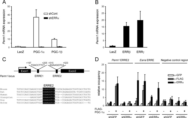
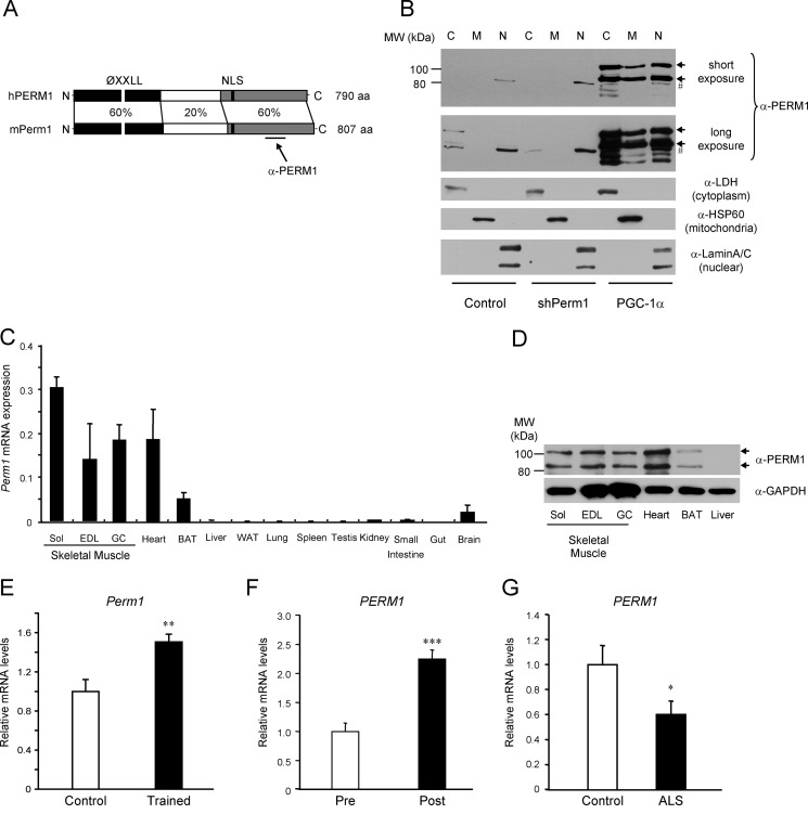
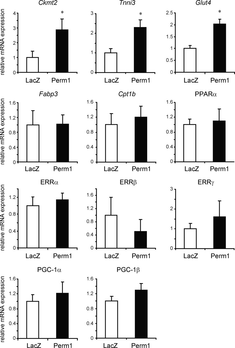
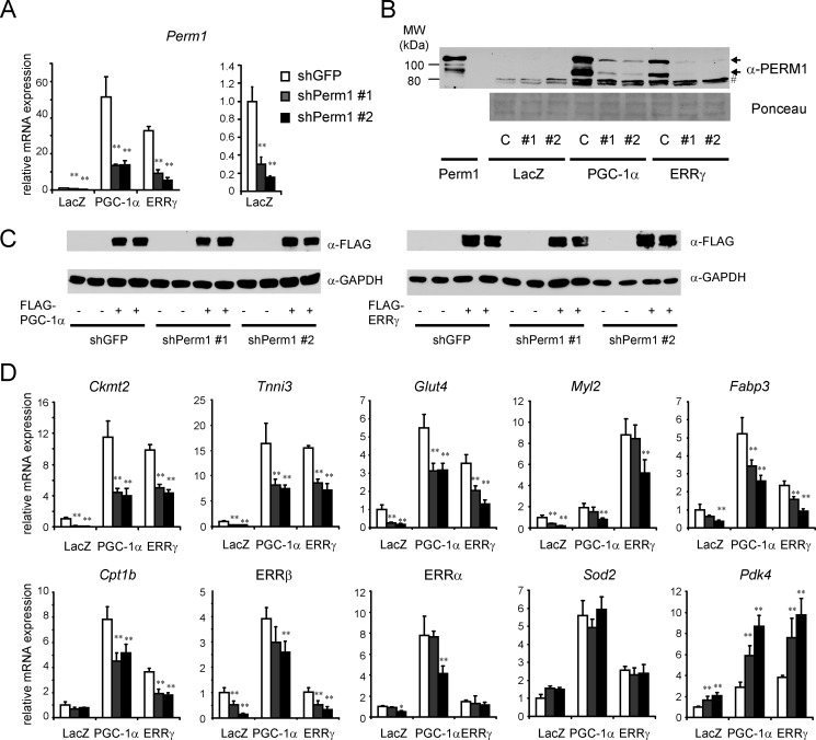
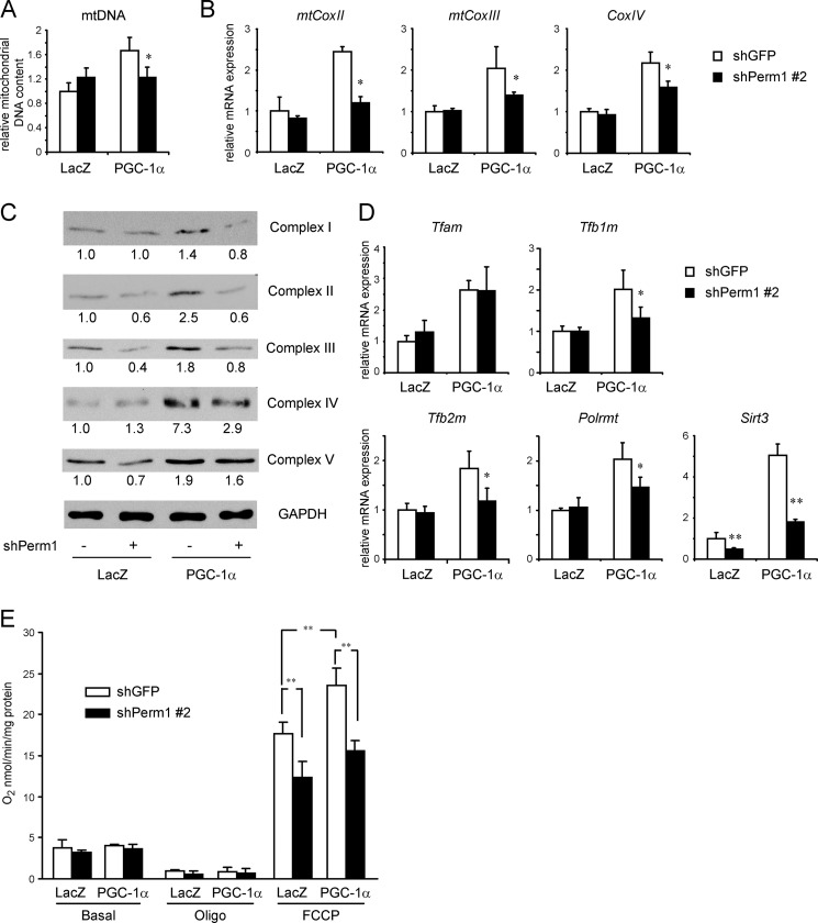
Mitochondrial oxidative metabolism and energy transduction pathways are critical for skeletal and cardiac muscle function. The expression of genes important for mitochondrial biogenesis and oxidative metabolism are under the control of members of the peroxisome proliferator-activated receptor γ coactivator 1 (PGC-1) family of transcriptional coactivators and the estrogen-related receptor (ERR) subfamily of nuclear receptors. Perturbations in PGC-1 and/or ERR activities have been associated with alterations in capacity for endurance exercise, rates of muscle atrophy, and cardiac function. The mechanism(s) by which PGC-1 and ERR proteins regulate muscle-specific transcriptional programs is not fully understood. We show here that PGC-1α and ERRs induce the expression of a so far uncharacterized muscle-specific protein, PGC-1- and ERR-induced regulator in muscle 1 (Perm1), which regulates the expression of selective PGC-1/ERR target genes. Perm1 is required for the basal as well as PGC-1α-enhanced expression of genes with roles in glucose and lipid metabolism, energy transfer, and contractile function. Silencing of Perm1 in cultured myotubes compromises respiratory capacity and diminishes PGC-1α-induced mitochondrial biogenesis. Our findings support a role for Perm1 acting downstream of PGC-1α and ERRs to regulate muscle-specific pathways important for energy metabolism and contractile function. Elucidating the function of Perm1 may enable novel approaches for the treatment of disorders with compromised skeletal muscle bioenergetics, such as mitochondrial myopathies and age-related/disease-associated muscle atrophies.

Keywords: Bioenergetics; Energy Metabolism; Estrogen-related Receptor; Mitochondrial Biogenesis; Nuclear Receptors; PGC-1α; Skeletal Muscle; Transcription Regulation.


Figures

FIGURE 1.
FIGURE 1.
PGC-1α, PGC-1β, and ERRs regulate Perm1 expression in myotubes. A, C2C12 myotubes were infected with adenoviruses expressing shGFP (shCont) or shERRα on day 4 of differentiation and LacZ, PGC-1α, or PGC-1β on day 6. RNA was harvested on day 7. Perm1 mRNA levels were determined by RT-qPCR, normalized to 36B4 levels in each sample, and expressed relative to Perm1 levels in control cells (LacZ/shCont). B, C2C12 myotubes were infected with adenoviruses expressing LacZ, ERRβ, or ERRγ on day 5. RNA was harvested on day 6, and Perm1 levels were quantified as in A. C, graphic representation of the Perm1 locus showing the location of the ERREs relative to the transcriptional start site and an alignment of the ERRE2 sequence (highlighted in black) across species. D, ChIPs were performed using antibodies against GFP (α-GFP; control), FLAG-tagged PGC-1α (α-FLAG), or ERRα (α-ERRα), and C2C12 myotubes infected with adenoviruses as described in A. The abundance of the Perm1 ERRE2, the Esrra ERRE (positive control for comparison), and a negative control genomic region in the ChIPs was quantified by qPCR, normalized to input signal, and expressed relative to the levels of each region in the control α-GFP samples. In A, B, and D, data are the mean of three to four experimental replicates from one of at least two representative experiments. Error bars represent S.D.
FIGURE 2.
FIGURE 2.
Perm1 encodes a protein found in multiple cellular compartments, is selectively expressed in muscle, and is regulated by physical activity. A, schematic representation of the human (h) and mouse (m) PERM1 proteins, showing the percent identity at the amino acid level and highlighting the conserved motifs (ØXXLL, protein interaction motif; NLS, nuclear localization signal; aa, amino acids) and the region recognized by the anti-PERM1 serum. B, detection of endogenous Perm1 in C2C12 myotubes infected with adenoviruses expressing shGFP (control), shPerm1, or PGC-1α on day 6 of differentiation. Cells were harvested on day 8, cell lysates were subjected to subcellular fractionation, and Perm1 protein was detected using the PERM1 antibody (α-PERM1). Endogenous Perm1 in control cells was detectable only after long exposure (second panel from top). Antibodies against cytoplasmic lactate dehydrogenase (LDH), mitochondrial HSP60, and nuclear lamins (three bottom panels) were used to assess the purity of the cytoplasmic (C), mitochondrial (M), and nuclear (N) fractions. Arrows indicate the two major protein isoforms encoded by Perm1; # indicates a nonspecific protein reacting with the antibody and enriched in the nuclear fraction. C, Perm1 mRNA levels in the indicated tissues of 10-week-old male mice (n = 3) were determined by RT-qPCR and normalized to levels of 36B4 in each tissue (Sol, soleus; EDL, extensor digitorum longus; GC, gastrocnemius). D, Perm1 protein in lysates from skeletal muscles, heart, brown adipose tissue (BAT), and liver of 10-week-old male mice was detected by SDS-PAGE and Western blot using the PERM1 antibody (upper panel); an antibody against GAPDH was used as a loading control (lower panel). E, Perm1 mRNA levels in the quadriceps muscles of 19-week-old female mice that had access to electronically monitored in-cage running wheels for 5 weeks (Trained; n = 7) and control sedentary littermate females (Control; n = 7) were determined by RT-qPCR, normalized to levels of GAPDH, and expressed relative to Perm1 levels in the control sedentary mice. F, PERM1 mRNA levels in muscle biopsies taken from male subjects (n = 7) before (Pre) or 3 h after (Post) an acute cycling bout (60 min at ∼70% of their VO2 peak) were quantified by RT-qPCR as described (41). G, PERM1 mRNA levels in biopsies taken from male control subjects or ALS patients (n = 9) were quantified by RT-qPCR as described (24). Error bars represent S.E. *, p < 0.05; **, p < 0.01; ***, p < 0.001.
FIGURE 3.
FIGURE 3.
Overexpressed Perm1 up-regulates selective PGC-1α/ERR target genes in myotubes. C2C12 myotubes were infected on day 4 of differentiation with adenoviruses expressing LacZ (control) or Perm1. RNA was harvested on day 6. RNA levels for the indicated genes were determined by RT-qPCR, normalized to 36B4 levels, and expressed relative to the levels of each gene in control (LacZ) cells. Data are the mean of four experimental replicates from one of at least two representative experiments. Error bars represent S.D. *, p < 0.05.
FIGURE 4.
FIGURE 4.
Endogenous Perm1 is required for the expression and/or induction of selective PGC-1α/ERR target genes. C2C12 myotubes were infected on day 4 of differentiation with adenoviruses expressing control (shGFP) or shPerm1 and on day 6 with adenoviruses expressing LacZ, PGC-1α, or ERRγ. RNA and protein were harvested on day 7. A and D, mRNA levels of Perm1 and the indicated PGC-1/ERR-regulated genes were measured by RT-qPCR, normalized to 36B4 levels, and expressed relative to levels of each gene in control cells (shGFP/LacZ). Perm1 mRNA levels in LacZ (control) cells are shown twice: in the same scale as in PGC-1α/ERRγ expressing cells (A, left panel) and in a smaller scale for better view of the mRNA levels (A, right panel). Data are the mean of three to four experimental replicates from one of at least two representative experiments. Error bars represent S.D. *, p < 0.05; **, p < 0.01. B, Perm1 protein levels were determined by Western blot using the PERM1 antibody. C, shGFP; #1, shPerm1-1; #2, shPerm1-2. The two arrows indicate the Perm1 protein isoforms. As a control, the first lane shows the Perm1 protein expressed from the cloned cDNA (myotubes infected with adenovirus expressing Perm1); the major protein species co-migrates with the upper isoform of the doublet seen for the endogenous protein. Even when expressed from the cloned cDNA, a faster migrating species is also detectable. Protein loading was assessed by Ponceau staining of the membrane (lower panel); lane 1 (lysates of C2C12 infected with Ad/Perm1) has 8-fold less protein than all other lanes). # indicates nonspecific bands. D, the levels of FLAG-tagged PGC-1α and FLAG-tagged ERRγ were determined by Western blot analysis using an anti-FLAG antibody (upper panel). As a control for protein loading, the same blots were probed with an anti-GAPDH antibody (lower panel).
FIGURE 5.
FIGURE 5.
Perm1 is required for PGC-1α-induced mitochondrial biogenesis and maximal oxidative capacity. C2C12 myotubes were infected on day 4 of differentiation with adenoviruses expressing control (shGFP) or shPerm1 and on day 6 with adenoviruses expressing LacZ or PGC-1α. DNA, RNA, and protein were harvested on day 7. A, the relative mitochondrial DNA content was determined as the ratio of mitochondrial (CoxII) DNA to genomic (Nrip1) DNA copy numbers and expressed relative to the ratio seen in control (shGFP/LacZ) myotubes. B, mRNA levels for the indicated mitochondrial and nuclear DNA-encoded OxPhos genes were determined by RT-qPCR, normalized to 36B4 levels, and expressed relative to levels of each gene in control (shGFP/LacZ) myotubes. C, the levels of OxPhos complexes were measured by Western blot analysis using total protein lysates and the Total OxPhos Complex antibody mixture. The intensity of the bands was quantified using ImageJ software. The values are expressed relative to the signal intensity in control (shGFP/LacZ) myotubes. D, mRNA levels of genes with roles in mitochondrial biogenesis were determined and expressed as in B. In A, B, and D, data are the mean of three to four experimental replicates from one of at least two representative experiments. Error bars represent S.D. *, p < 0.05; **, p < 0.01. E, oxygen consumption rates of the myotubes were analyzed on day 7 in the absence (Basal) or presence of 2.5 μg/ml oligomycin (Oligo) and a 1.6 μm concentration of the uncoupling agent FCCP. Rates are normalized to the protein content of the cells and are the mean of three experimental replicates from one of two representative experiments. Error bars represent S.D. **, p < 0.01.

References

    1. Wallace D. C. (2000) Mitochondrial defects in cardiomyopathy and neuromuscular disease. Am. Heart J. 139, S70–S85 - PubMed
    1. DiMauro S. (2006) Mitochondrial myopathies. Curr. Opin. Rheumatol. 18, 636–641 - PubMed
    1. van Adel B. A., Tarnopolsky M. A. (2009) Metabolic myopathies: update 2009. J. Clin. Neuromuscul. Dis. 10, 97–121 - PubMed
    1. Ingwall J. S. (2009) On the control of metabolic remodeling in mitochondria of the failing heart. Circ. Heart Fail. 2, 275–277 - PubMed
    1. Rowe G. C., Jiang A., Arany Z. (2010) PGC-1 coactivators in cardiac development and disease. Circ. Res. 107, 825–838 - PMC - PubMed

Publication types

MeSH terms

Substances

Associated data