Review
. 2013 Oct;14(10):1029-41.
doi: 10.1111/tra.12091. Epub 2013 Jul 30.

Hidden behind autophagy: the unconventional roles of ATG proteins

Affiliations
Review

Hidden behind autophagy: the unconventional roles of ATG proteins

Jovanka Bestebroer et al. Traffic. 2013 Oct.

Abstract

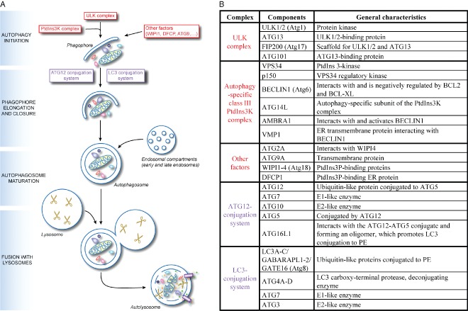
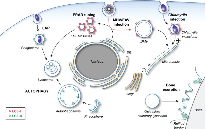
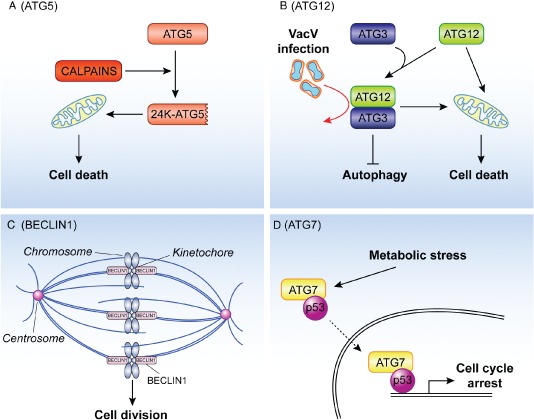
Macroautophagy (hereafter referred to as autophagy) is an evolutionarily conserved intracellular catabolic transport route that generally allows the lysosomal degradation of cytoplasmic components, including bulk cytosol, protein aggregates, damaged or superfluous organelles and invading microbes. Target structures are sequestered by double-membrane vesicles called autophagosomes, which are formed through the concerted action of the autophagy (ATG)-related proteins. Until recently it was assumed that ATG proteins were exclusively involved in autophagy. A growing number of studies, however, have attributed functions to some of them that are distinct from their classical role in autophagosome biogenesis. Autophagy-independent roles of the ATG proteins include the maintenance of cellular homeostasis and resistance to pathogens. For example, they assist and enhance the turnover of dead cells and microbes upon their phagocytic engulfment, and inhibit murine norovirus replication. Moreover, bone resorption by osteoclasts, innate immune regulation triggered by cytoplasmic DNA and the ER-associated degradation regulation all have in common the requirement of a subset of ATG proteins. Microorganisms such as coronaviruses, Chlamydia trachomatis or Brucella abortus have even evolved ways to manipulate autophagy-independent functions of ATG proteins in order to ensure the completion of their intracellular life cycle. Taken together these novel mechanisms add to the repertoire of functions and extend the number of cellular processes involving the ATG proteins.

Keywords: ATG proteins; apoptosis; autophagy; degradation; immunity; infection; pathogens; subversion; unconventional.


Figures

Figure 1
Figure 1
The mechanism of canonical autophagy and the conventional functions of the ATG proteins. A) The mechanism of autophagy. Upon induction, the early steps of autophagosome biogenesis entail the formation of the phagophore through the orchestrated action of the ULK complex, the PtdIns3K complex and other factors including ATG9, VMP1, AMBRA1, DFCP1 and the WIPI proteins. Subsequently the ATG12 and LC3 conjugation systems are key in mediating the expansion and nucleation of the phagophore into an autophagosome, an event that allows the sequestration of the cargo targeted for degradation. Once complete, autophagosomes first fuse with endosomal compartments to mature into amphisomes before fusing with lysosomes to form autolysosomes. Resident hydrolases (indicated with scissors) consume the autophagosomal cargo and internal vesicles into metabolites (amino acids, sugars, nucleotides and other basic molecules), which are subsequently transported into the cytoplasm by permeases and possibly other transporters (depicted as small violet pipes), where they are used as either a source of energy or as building blocks for the synthesis of new macromolecules. B) The key autophagy proteins and their organization in functional gene clusters. The ULK complex and the autophagy‐specific class III PtdIns3K complex, plus other factors, are involved in the initiation of autophagosome biogenesis (highlighted in red in panel A) whereas the ATG12‐ and LC3‐conjugation systems are mostly involved in the phagophore elongation (highlighted in violet in panel A). While the yeast homologues of the ATG2A, ATG9A and WIPI are interacting, there is no evidence this also holds true for their mammalian counterparts. Moreover, there are no indications that DFCP1 associate with these components. The yeast homologs of the mammalian ATGs proteins are indicated in between brackets. Adapted from 4.
Figure 2
Figure 2
The cellular functions of LC3. After post‐translational processing by ATG4, LC3‐I is covalently linked to PE to generate LC3‐II through the action of two ubiquitin‐like conjugation systems. The formation of lipidated LC3 is essential for autophagosome formation by mediating the expansion of the phagophore. LC3‐II is also involved in LAP as well as the specialized secretion by osteoclasts, which is essential for the establishment of the ruffled borders for bone resorption. The role of LC3‐II in these two latter pathways is unclear, but its localization and function do not require an intact ATG machinery. Precursor LC3‐I is also carrying out cellular functions, which do not appear to involve most of the other ATG proteins. LC3‐I is recruited together with MAP1 proteins onto Chlamydia inclusions and allows the redistribution of these bacteria‐containing compartments in proximity of the Golgi through its interaction with the microtubules. LC3‐I is also associated with the surface of the EDEMosomes via its interaction with the protein cargo receptor Sel1L. The role of LC3‐I in the ERAD tuning pathway remains unknown, but it could act as an adaptor for either a protein vesicle coat or the cytoskeleton network. Viruses, such as MHV and EAV, hijack this transport route to generate their replicative DMVs, which are also decorated with LC3‐I (red arrow). LC3‐I is essential for both the ERAD tuning and the replication of these viruses. In this latter context, LC3‐I could mediate the microtubule‐dependent subcellular redistribution of DMVs.
Figure 3
Figure 3
The unconventional roles of ATG proteins in cell death and cell division and proliferation. A) Apoptotic stimuli activate calpains and one of the substrates of these proteases is ATG5. The resulting 24 kDa truncated form of ATG5 (24K‐ATG5) associates with mitochondria and triggers cytochrome c release through a still unknown mechanism, thus participating to the initiation of the cell death programs. B) ATG12 participates in apoptosis at least in two distinct ways. First, it can become part of the ATG12‐ATG3 conjugate, which plays an important role in controlling mitochondria homeostasis and sensitizes cells to apoptosis triggered by the intrinsic mitochondrial pathway. The ATG12‐ATG3 conjugate is also stimulated upon vaccinia virus infection and it is required for the life cycle of this virus (red arrow). Second and independently from its capacity to be covalently linked to ATG5 or ATG3, ATG12 stimulates mitochondrial apoptosis through its binding and inhibition of the anti‐apoptotic function of the members of the BCL‐2 protein family. C) During mitosis BECLIN1 associates with ZWINT‐1 and mediates the interaction between the kinetochore and microtubules, a key event in cell division. Depletion of BECLIN1 causes a defect in the kinetochore protein assembling, misalignment of chromosomes and a general block in mitotic progression. D) Upon metabolic stress, ATG7 is found associated with p53 in the cytoplasm and nucleus. The nuclear complex positively regulates the expression of genes mediating the cell cycle arrest. It remains to be determined whether the nuclear ATG7‐p53 pool results from the translocation into the nucleus of the subpopulation of this complex formed in the cytoplasm (dashed arrow).

References

    1. Kraft C, Martens S. Mechanisms and regulation of autophagosome formation. Curr Opin Cell Biol 2012;24:496–501. - PubMed
    1. Yang Z, Klionsky DJ. Mammalian autophagy: core molecular machinery and signaling regulation. Curr Opin Cell Biol 2010;22:124–131. - PMC - PubMed
    1. Kroemer G, Marino G, Levine B. Autophagy and the integrated stress response. Mol Cell 2010;40:280–293. - PMC - PubMed
    1. Levine B, Mizushima N, Virgin HW. Autophagy in immunity and inflammation. Nature 2011;469:323–335. - PMC - PubMed
    1. Choi AM, Ryter SW, Levine B. Autophagy in human health and disease. N Engl J Med 2013;368:651–662. - PubMed

Publication types

LinkOut - more resources