. 2013 Jul 9;15(4):R73.
doi: 10.1186/ar4250.

Modulation of monosodium urate crystal-induced responses in neutrophils by the myeloid inhibitory C-type lectin-like receptor: potential therapeutic implications

Modulation of monosodium urate crystal-induced responses in neutrophils by the myeloid inhibitory C-type lectin-like receptor: potential therapeutic implications

Valérie Gagné et al. Arthritis Res Ther. .

Abstract

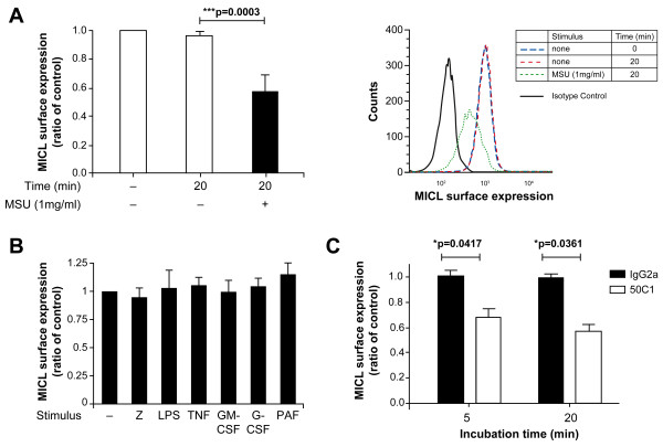
Introduction: Monosodium urate crystals (MSU), the etiological agent of gout, are one of the most potent proinflammatory stimuli for neutrophils. The modulation of MSU-induced neutrophil activation by inhibitory receptors remains poorly characterized. The expression of the myeloid inhibitory C-type lectin-like receptor (MICL) in neutrophils is downregulated by several proinflammatory stimuli, suggestive of a role for this receptor in neutrophil function. We thus investigated the potential role of MICL in MSU-induced neutrophil activation.

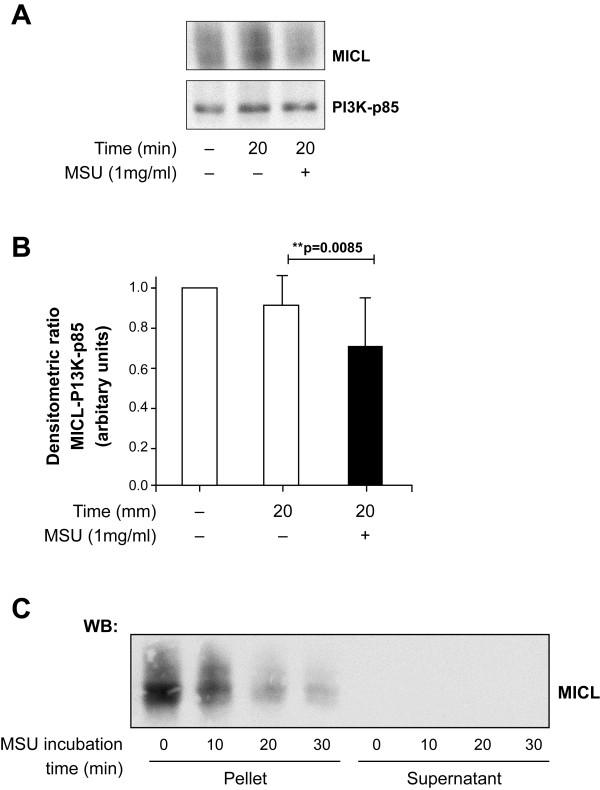
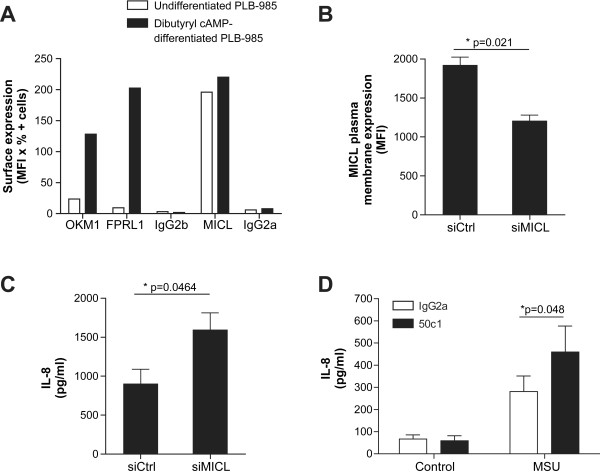
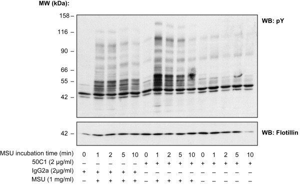
Methods: The expression of MICL was monitored in human neutrophils by flow cytometry and Western blot analysis after stimulation with MSU. Protein tyrosine phosphorylation was also assessed by Western blot analysis and the production of IL-1 and IL-8 by enzyme-linked immunosorbent assay. Changes in the concentration of cytoplasmic free calcium were monitored with the Fura-2-acetoxymethyl ester calcium indicator. MICL expression was modulated with an anti-MICL antibody in neutrophils and siRNA in the PLB-985 neutrophil-like cell line.

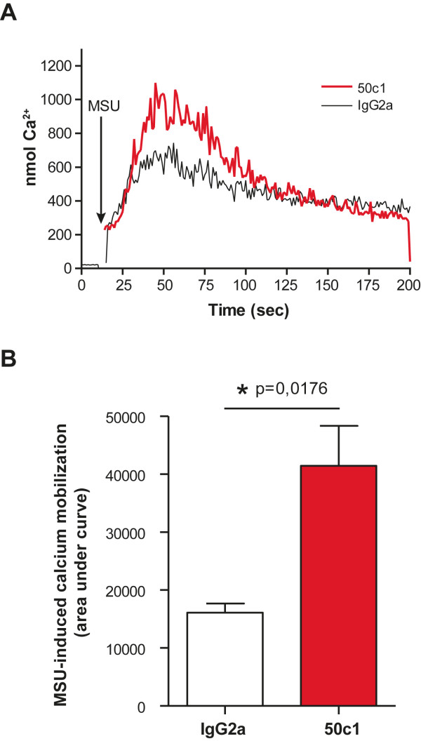
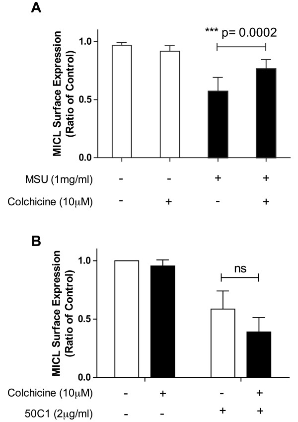
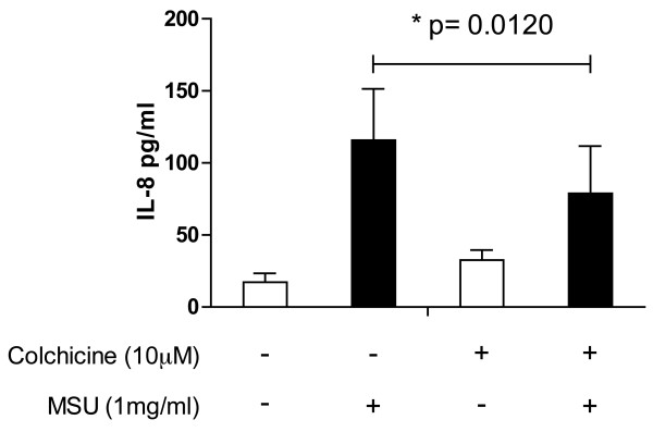
Results: MSU induced the downregulation of MICL expression in neutrophils. A diminution in the expression of MICL induced by antibody cross-linking or siRNA enhanced the MSU-dependent increase in cytoplasmic calcium levels, protein tyrosine phosphorylation and IL-8 but not IL-1 production. Pretreatment of neutrophils with colchicine inhibited the MSU-induced downregulation of MICL expression.

Conclusions: Our findings strongly suggest that MICL acts as an inhibitory receptor in human neutrophils since the downregulation of MICL expression enhances MSU-induced neutrophil activation. Since MSU downregulates the expression of MICL, MICL may play a pathogenic role in gout by enhancing neutrophil effector functions. In support of this notion, colchicine counteracts the MSU-induced loss of MICL expression. Our findings thus also provide further insight into the potential molecular mechanisms behind the anti-inflammatory properties of this drug.


Figures

Figure 1
Figure 1
Surface myeloid inhibitory C-type lectin-like receptor (MICL) expression is significantly decreased upon activation of human neutrophils with monosodium urate crystals (MSU). The plasma membrane expression of MICL was examined by flow cytometry on freshly isolated neutrophils (10 × 106 cells/ml) after incubation at 37°C with (A) MSU (1 mg/ml) for 20 min, (B) nonopsonized zymosan (z) (ratio = 10 z/cell), lipopolysaccharide (LPS) (22.5 ng/ml in 1× Hanks' balanced salt solution containing 5% decomplemented fetal bovine serum), tumor necrosis factor α (TNF-α; 100 U/ml), granulocyte-macrophage colony-stimulating factor (80 nM), granulocyte colony-stimulating factor (50 ng/ml) or platelet-activating factor (10−6 M) for 15 min or (C) the 50C1 antibody (1 µg/ml) or IgG2a isotype control antibody for 5 min or 20 min as described in Methods. MICL expression was compared to the control. The raw flow cytometry data in (A) is shown in the right panel. These graphs are compilations from four independent experiments.
Figure 2
Figure 2
Total MICL expression is reduced upon activation of human neutrophils with MSU. (A) Freshly isolated human neutrophils (20 × 106 cells/ml) were incubated with MSU (1 mg/ml) at 37°C, then the stimulation was terminated. Aliquots of the suspension were stopped at 20 min by transferring it directly into the same volume of nonreducing 2× boiling modified Laemmli sample buffer. Whole-cell lysates were probed by Western blotting for MICL (anti-MICL clone HB3; upper panel) and phosphoinositide 3-kinase (PI3K)-p85 (lower panel) as loading control. This result is representative of eight independent experiments. (B) Densitometric ratios from (A) are represented on the graph. (C) Human neutrophils (20 × 106 cells/ml) were incubated with MSU (1 mg/ml) at 37°C. Aliquots of the suspension were centrifuged, and pellets were stopped at the indicated times in nonreducing 2× boiling modified Laemmli sample buffer. The supernatants were precipitated with 2,2,2-trichloroacetic acid as described in Methods. Pellets and supernatants were probed by Western blotting for MICL (anti-MICL clone HB3). This result is representative of three independent experiments.
Figure 3
Figure 3
MICL modulates MSU-induced IL-8 secretion in human neutrophils. The PLB-985 cell line was differentiated with dibutyryl cyclic adenosine monophosphate (dibutyryl cAMP) for 72 h as described in Methods. The plasma membrane expression of Mac-1 (OKM1), the formyl peptide receptor-like 1 (FPRL1) and MICL were analyzed by flow cytometry on differentiated and undifferentiated PLB-985 cells. Immunoglobulin G2b (IgG2b) and IgG2a were used as control isotypes. (B) Dibutyryl cAMP-differentiated, neutrophil-like PLB-985 (20 × 106 cells/ml) were transfected using a nucleofection system with a MICL-specific small interfering RNA (siMICL) or with a negative control small interfering RNA (siCtrl). Cells were then stimulated for 3 h at 37°C with 1 mg/ml MSU in RPMI 1640 medium. Cells were centrifuged (16,000 × g for 5 min), and the supernatants were harvested and filtered. Extracellular interleukin 8 (IL-8) was quantitated using commercially available enzyme-linked immunosorbent assay (ELISA) kits. All samples were measured in duplicate. This graph is a compilation of four independent experiments. (C) MICL surface expression was monitored by flow cytometry following siRNA transfections. Cells (10 × 106 cells/ml) were incubated with 50C1 antibody (2 µg/ml) for 30 min on ice, washed and incubated with fluorescein isothiocyanate (FITC)-labeled goat antimouse Fcγ-specific IgG (diluted 1:100 in Hanks' balanced salt solution/bovine serum albumin (HBSS/BSA)) for 30 min on ice. Cells were then washed twice in HBSS/BSA and analyzed by flow cytometry using a BD FACSCanto II flow cytometer obtained from BD Biosciences. This graph is a compilation of four independent experiments. (D) Human neutrophils (20 × 106 cells/ml) were incubated with anti-MICL antibody (clone 50C1, 2 μg/ml) or IgG2a isotype for 5 min at 37ºC and then washed. Cells were then stimulated with MSU at 1 mg/ml or not for 3 h at 37°C. Extracellular IL-8 was monitored as described in (B). All samples were measured in triplicate. This result is a compilation of five independent experiments.
Figure 4
Figure 4
MICL does not modulate MSU-induced IL-1β production in primed, human neutrophils. Resting human neutrophils (20 × 106 cells/ml) or neutrophils incubated with the IgG2a isotype antibody, 50C1, in the presence or absence of TNF-α were stimulated with MSU (1 mg/ml) for 8 h at 37°C and centrifuged, and IL-1β was quantified in the cell-free supernatant by ELISA as described in Methods. All samples were measured in duplicate. This graph is a compilation of four independent experiments.
Figure 5
Figure 5
MICL modulates the MSU-induced tyrosine phosphorylation pattern in human neutrophils. Human neutrophils (20 × 106 cells/ml) were preincubated with anti-MICL antibody (clone 50C1, 2 μg/ml) (+) or IgG2a isotype (-) for 5 min and then washed. Next, cells were stimulated with MSU (1 mg/ml) at 37°C. Aliquots of the cell suspension were stopped at the indicated times by transferring them directly into the same volume of 2× boiling modified Laemmli sample buffer. Whole-cell lysates were probed by Western blotting for tyrosine phosphorylation (clone 4G10, upper panel) and flotillin-1 (lower panel), the loading control. This result is representative of three independent experiments.
Figure 6
Figure 6
MICL modulates MSU-induced calcium mobilization in human neutrophils. (A) Human neutrophils (10 × 106 cells/ml) were preincubated with Fura-2AM (1 µM) for 30 min at 37°C. The extracellular probe was removed by washing in HBSS, and the cells were resuspended at 5 × 10⁶ cells/ml. Anti-MICL antibody (clone 50C1, 2 μg/ml) or IgG2a isotype was added for 5 min. Cells were transferred to a thermostat-controlled (37°C) cuvette compartment of an SLM 8000 spectrofluorometer. Calcium mobilization was measured following the addition of 1 mg/ml MSU at 37°C. Fluorescence was monitored at an excitation wavelength of 340 nm and an emission wavelength of 510 nm. The internal calcium concentrations were calculated as described by Grynkiewicz et al. [16]. This result is representative of four independent experiments.
Figure 7
Figure 7
Colchicine inhibits the MSU-induced internalization of MICL in human neutrophils. Freshly isolated human neutrophils were treated with colchicine (10 µM) (+) or dimethyl sulfoxide (DMSO) (-) for 30 min at 37°C and then incubated with (A) MSU (1 mg/ml) for 20 min at 37°C or (B) 50C1 for 5 min at 37°C. The stimulations were terminated by transferring the tubes to an ice bath, followed by centrifugation at 400 × g for 2 min at 4°C. The cell pellets were washed in cold HBSS containing 0.005% BSA and incubated with (A) 50C1 for 30 min, followed by incubation with FITC-labeled goat antimouse Fcγ-specific IgG (diluted 1:100 in HBSS/BSA) for 30 min on ice, or (B) FITC-labeled goat anti-mouse Fcγ-specific IgG (diluted 1:100 in HBSS/BSA) for 30 min on ice. Cells were then washed twice in HBSS/BSA and analyzed by flow cytometry using the FACSCanto II flow cytometer (BD Biosciences). This graph is a compilation of 7 independent experiments in (A) and 3 independent experiments in (B).
Figure 8
Figure 8
Colchicine modulates MSU-induced IL-8 secretion in human neutrophils. Neutrophils were incubated in colchicine (10 µM) (+) or DMSO (-) for 30 min at 37°C prior to adding MSU (1 mg/ml) or buffer to the neutrophil-colchicine mixture and incubating it for a further 3 h at 37°C. The quantity of IL-8 in the supernatant was determined by ELISA. A compilation of the data from three independent experiments is shown (n = 3).

Comment in

Similar articles

Cited by

References

    1. Martinon F. Mechanisms of uric acid crystal-mediated autoinflammation. Immunol Rev. 2010;15:218–232. doi: 10.1111/j.0105-2896.2009.00860.x. - DOI - PubMed
    1. Weaver AL. Epidemiology of gout. Cleve Clin J Med. 2008;15(Suppl 5):S9–S12. - PubMed
    1. Busso N, So A. Mechanisms of inflammation in gout. Arthritis Res Ther. 2010;15:206. doi: 10.1186/ar2952. - DOI - PMC - PubMed
    1. Popa-Nita O, Naccache PH. Crystal-induced neutrophil activation. Immunol Cell Biol. 2010;15:32–40. doi: 10.1038/icb.2009.98. - DOI - PubMed
    1. Daëron M, Jaeger S, Du Pasquier L, Vivier E. Immunoreceptor tyrosine-based inhibition motifs: a quest in the past and future. Immunol Rev. 2008;15:11–43. doi: 10.1111/j.1600-065X.2008.00666.x. - DOI - PubMed

Publication types