. 2013 Oct;17(10):1247-60.
doi: 10.1111/jcmm.12100. Epub 2013 Jul 9.

Human endometrial stem cells confer enhanced myocardial salvage and regeneration by paracrine mechanisms

Affiliations

Human endometrial stem cells confer enhanced myocardial salvage and regeneration by paracrine mechanisms

Zhi Jiang et al. J Cell Mol Med. 2013 Oct.

Abstract

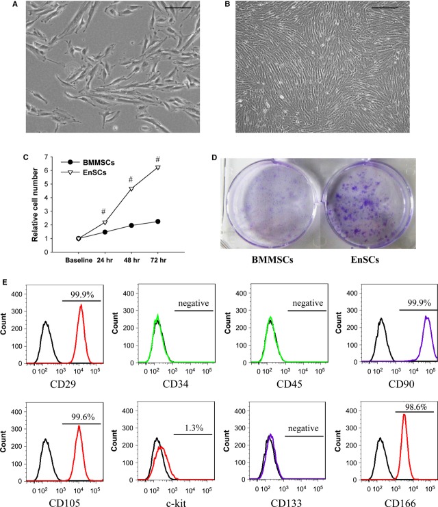
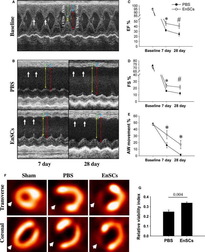
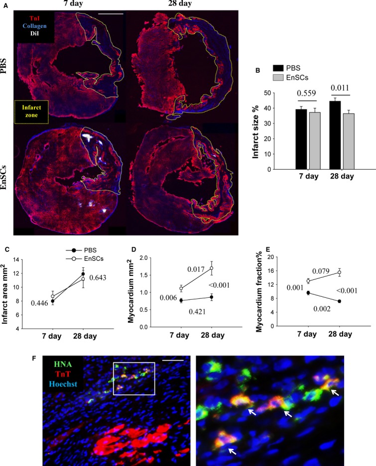
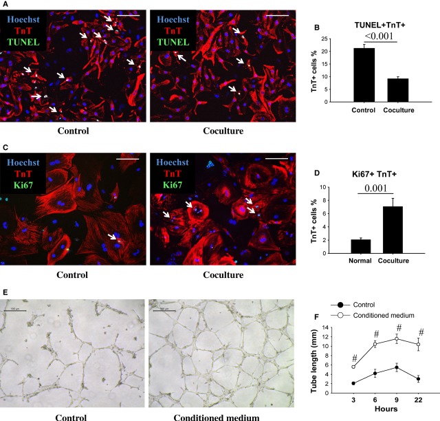
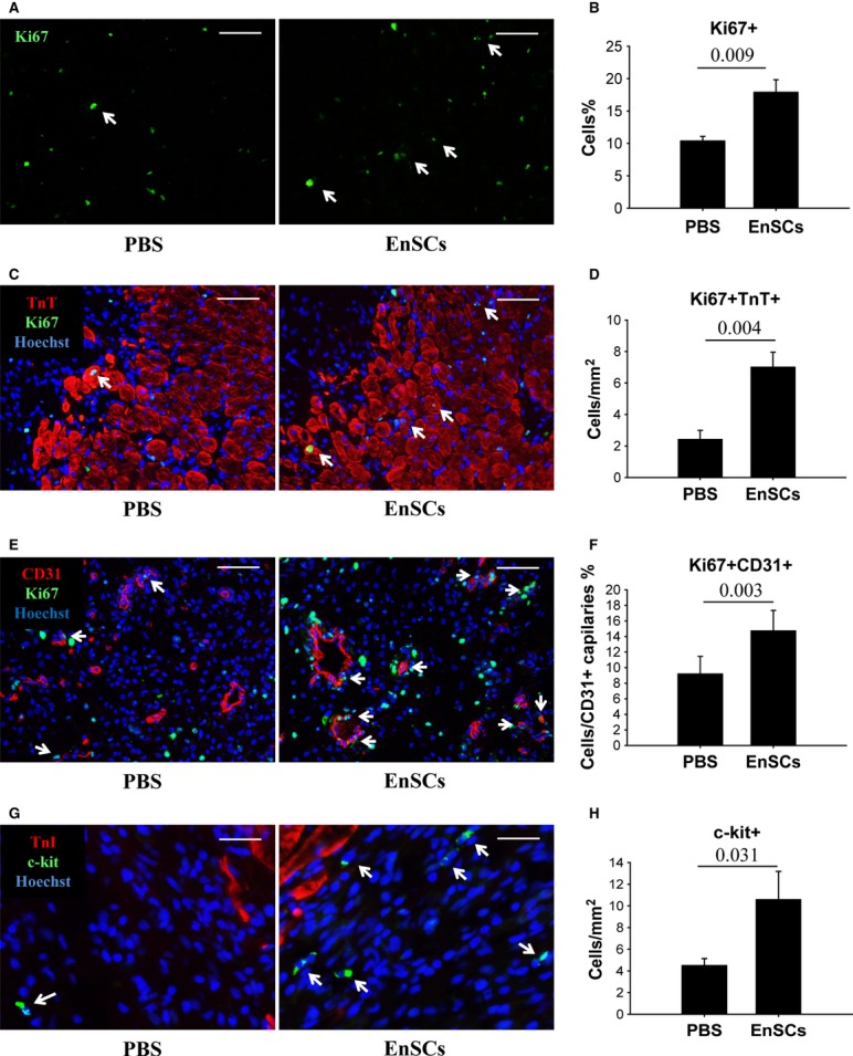
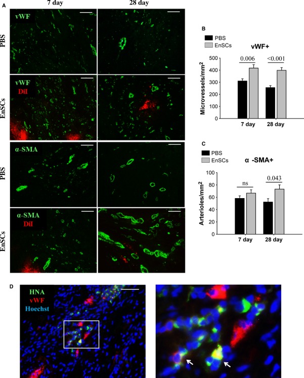
Human endometrial stem cells (EnSCs) have the potential to be 'off the shelf' clinical reagents for the treatment of heart failure. Here, using an immunocompetent rat model of myocardial infarction (MI), we provide evidence that the functional benefits of EnSC transplantation are principally and possibly exclusively through a paracrine effect. Human EnSCs were delivered by intramyocardial injection into rats 30 min. after coronary ligation. EnSC therapy significantly preserved viable myocardium in the infarct zone and improved cardiac function at 28 days. Despite increased viable myocardium and vascular density, there was scant evidence of differentiation of EnSCs into any cardiovascular cell type. Cultured human EnSCs expressed a distinctive profile of cytokines that enhanced the survival, proliferation and function of endothelial cells in vitro. When injected into the peri-infarct zone, human EnSCs activated AKT, ERK1/2 and STAT3 and inhibited the p38 signalling pathway. EnSC therapy decreased apoptosis and promoted cell proliferation and c-kit+ cell recruitment in vivo. Myocardial protection and enhanced post-infarction regeneration by EnSCs is mediated primarily by paracrine effects conferred by secreted cytokines that activate survival pathways and recruit endogenous progenitor stem cells. Menstrual blood provides a potentially limitless source of biologically competent 'off the shelf' EnSCs for allogeneic myocardial regenerative medicine.

Keywords: angiogenesis; apoptosis; endometrial stem cells; myocardial infarction; paracrine; regeneration.


Figures

Fig. 1
Fig. 1
Morphology and phenotype of human EnSCs. (A) Phase-contrast microscopic view of normal cultured 30% confluent EnSCs, passage 9. Scale bar denotes 100 μm. (B) Phase-contrast microscopic view of normal cultured 100% confluent EnSCs, passage 9. Scale bar denotes 200 μm. (C) Quantification of relative cell number. Cell number measured at 24, 48 and 72 was standardized by baseline. #P < 0.01 versus BMMSCs. (D) Representative pictures of colony formation assay. (E) Flow cytometric analysis of cell surface marker on EnSCs. EnSCs showed mesenchymal characteristics.
Fig. 2
Fig. 2
Functional benefits after the transplantation of EnSCs. (A and B) Representative M-mode echocardiographic images. The anterior wall (AW) movement was slightly preserved in EnSC group. Coloured lines showed the measurement of AW thickness and LV diameter. (C and E) Quantitative analysis of echocardiography (n = 8–14/group at each time-point). EnSCs transplantation increased ejection fraction, fractional shortening and improved AW movement at both 7 and 28 days. *P < 0.05 versus PBS group; #P < 0.01 versus PBS group. (F) Representative microPET images at transverse and coronal section at 28 days. PBS group showed radioactive defects as compared with sham group, but EnSC group showed low and distinct radioactivity in the infarct zone (white arrows), indicating viable cardiomyocytes in the zone. (G) Quantification of viable myocardium. The relative viability index was calculated by the region of interest method (n = 4/group). P values are shown at the top of bars.
Fig. 3
Fig. 3
Transplantation of EnSCs preserved myocardium and enhanced myocardium regeneration, but few directly differentiated into cardiomyocytes in vivo. (A) Representative double staining of TnT in red and collagen type I in blue to show myocardium and scar at transverse section. Yellow dotted box labelled the infarct zone. Scale bar denotes 2 mm. (B and C) Quantification of infarct size and infarct area (n = 9–11/group at each time-point). The infarct size did not differ at 7 days, but was larger in PBS group than EnSC group at 28 days. P values are shown in the figures. (D and E) Quantification of myocardium area and myocardium fraction (n = 9–11/group at each time-point). The EnSC group had larger myocardium area and higher myocardium fraction than PBS group. The increased myocardial area in EnSC group from 7 to 28 days indicated a small proportion of myocardial regeneration. P values were shown in the figures. (F) Representative images of human nuclear antigen–positive and TnT-positive cells at 28 days (white arrows). Scale bar denotes 50 μm.
Fig. 4
Fig. 4
The cytokines from EnSCs enhanced cell survival, proliferation and tube formation in vitro. (A) Representative pictures of TUNEL-positive cardiomyocytes (white arrows). Scale bar denotes 100 μm. (B) Quantification of the apoptotic cardiomyocytes (n = 8/group). Coculture with EnSCs decreased the ratio of apoptotic cardiomyocytes as compared with control group. P values are shown at the top of bars. (C) Representative pictures of Ki67-positive cardiomyocytes (white arrows). Scale bar denotes 100 μm. (D) Quantification of proliferating cardiomyocytes in vitro (n = 9/group). Coculture with EnSCs increased the number of proliferating cardiomyocytes as compared with control group. P values are shown at the top of bars. (E) Representative pictures showed tube formation of HUVECs. Scale bar denotes 100 μm. (F) Quantitative analysis of the tube length (n = 4/group at each time-point). The EnSCs conditioned medium increased tube length as compared with control group. #P < 0.01 versus control group.
Fig. 5
Fig. 5
Transplantation of EnSCs reduced cell apoptosis in vivo. Rats transplanted with EnSCs were killed at 2 days. The hearts were collected for analysis. (A) Representative pictures of TUNEL-positive nuclei. Scale bar denotes 50 μm. (B) Quantification of the apoptotic nuclei (n = 5/group). EnSCs transplantation reduced apoptotic nuclei density in both infarct and border zone. P values are shown at the top of bars. (C) Representative picture of DNA products of PCR. Human-specific cytokine gene expression was detected from the hearts of EnSCs group. (D) Quantification of proteins detected by western blot (n = 3–5/group). Transplantation of EnSCs increased phosphorylation of AKT, ERK and STAT3, increased Bcl-xl expression and inhibited caspase3 cleavage. *P < 0.05 versus PBS group; #P < 0.01 versus PBS group.
Fig. 6
Fig. 6
Transplantation of EnSCs promoted cell proliferation and c-kit-positive cell recruitment in vivo. Rats transplanted with EnSCs were killed at 7 days. The hearts were collected for immunofluorescent staining. (A) Representative pictures of immunostaining of Ki67+ cells in the infarct border (white arrows indicated proliferating cardiomyocytes). Scale bar denotes 100 μm. (B) Quantification of Ki67+ cells. (C) Representative pictures of colocalizing Ki67+ with TnT+ cells to demonstrate the proliferating cardiomyocytes (white arrows). Scale bar denotes 100 μm. (D) Quantification of Ki67+ TnT+ cells (n = 5–6/group). (E) Representative pictures of colocalizing Ki67+ (green) with CD31+ cells (red) to demonstrate the proliferating endothelial cells (white arrows). Scale bar denotes 50 μm. (F) Quantification of Ki67+ CD31+ cells (n = 5–6/group). (G) Representative pictures of c-kit+ cells (white arrows). Scale bar denotes 25 μm. (H) Quantification of c-kit+ cells (n = 8–9/group). P values are shown at the top of bars.
Fig. 7
Fig. 7
Transplantation of EnSCs stimulated angiogenesis in vivo through paracrine effect. (A) Representative immunofluorescence staining of vWF-positive microvessels and α-SMA-positive arterioles. Scale bar denotes 100 μm. (B and C) Quantification of vWF-positive microvessel and α-SMA-positive arteriole density (n = 5–6/group at each time-point). The vWF-positive microvessel density was greatly increased in EnSC group at 7 and 28 days. Arteriole density did not differ at 7 days, but was higher at 28 days in EnSC group than PBS group. P values are shown at the top of bars. (D) Representative image of human nuclear antigen–positive and vWF-positive cells at 28 days (white arrows). Scale bar denotes 50 μm.

References

    1. Roger VL, Go AS, Lloyd-Jones DM, et al. Heart disease and stroke statistics—2012 update. Circulation. 2012;125:e2–220. - PMC - PubMed
    1. Beitnes JO, Lunde K, Brinchmann JE, et al. Stem cells for cardiac repair in acute myocardial infarction. Expert Rev Cardiovasc Ther. 2011;9:1015–25. - PubMed
    1. Choi YH, Kurtz A, Stamm C. Mesenchymal stem cells for cardiac cell therapy. Hum Gene Ther. 2011;22:3–17. - PubMed
    1. Malliaras K, Li T-S, Luthringer D, et al. Safety and efficacy of allogeneic cell therapy in infarcted rats transplanted with mismatched cardiosphere-derived cells/clinical perspective. Circulation. 2012;125:100–12. - PMC - PubMed
    1. Cui C-H, Uyama T, Miyado K, et al. Menstrual blood-derived cells confer human dystrophin expression in the murine model of duchenne muscular dystrophy via cell fusion and myogenic transdifferentiation. Mol Biol Cell. 2007;18:1586–94. - PMC - PubMed

Publication types