Review
. 2013 Jul;11(3):150-64.
doi: 10.1016/j.jtos.2012.10.004. Epub 2013 Feb 13.

Corneal gene therapy: basic science and translational perspective

Affiliations
Review

Corneal gene therapy: basic science and translational perspective

Rajiv R Mohan et al. Ocul Surf. 2013 Jul.

Abstract

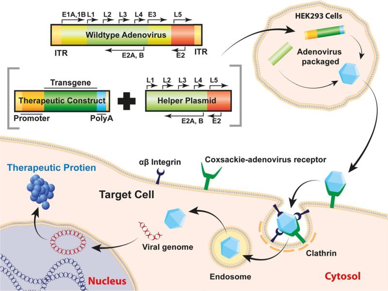
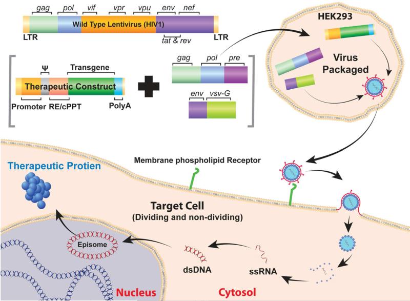
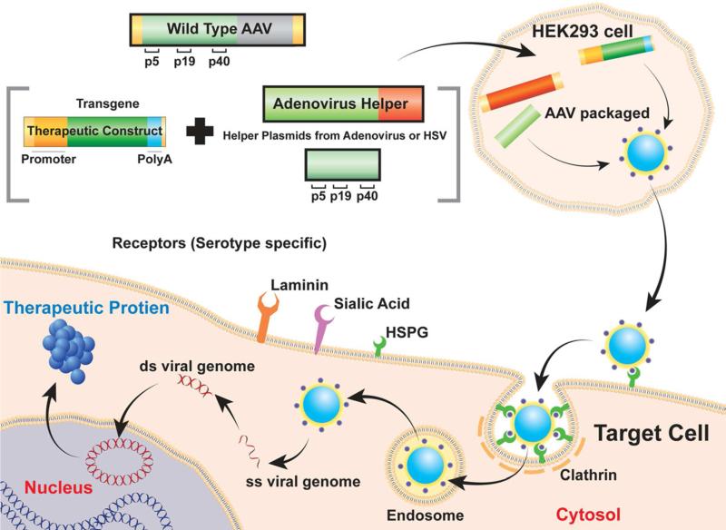
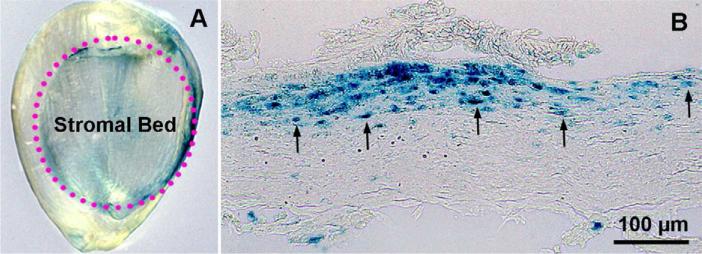
Corneal blindness is the third leading cause of blindness worldwide. Gene therapy is an emerging technology for corneal blindness due to the accessibility and immune-privileged nature of the cornea, ease of vector administration and visual monitoring, and ability to perform frequent noninvasive corneal assessment. Vision restoration by gene therapy is contingent upon vector and mode of therapeutic gene introduction into targeted cells/tissues. Numerous efficacious vectors, delivery techniques, and approaches have evolved in the last decade for developing gene-based interventions for corneal diseases. Maximizing the potential benefits of gene therapy requires efficient and sustained therapeutic gene expression in target cells, low toxicity, and a high safety profile. This review describes the basic science associated with many gene therapy vectors and the present progress of gene therapy carried out for various ocular surface disorders and diseases.

Keywords: adeno-associated virus; adenovirus; cornea; corneal diseases and dystrophies; gene therapy; lentivirus; nanoparticles; retrovirus.


Figures

Figure 1
Figure 1
Schematic illustration of adenovirus genome, mechanism of entry, and therapeutic gene production in host cell.
Figure 2
Figure 2
Schematic illustration of lentivirus genome, mechanism of entry, and therapeutic gene production in host cell.
Figure 3
Figure 3
Representative montage of the reconstructed mouse corneas showing actual surface area, quantity, and location of transgene in the entire cornea detected at two weeks. The transgene was delivered in vivo with lentivirus vector encoding for green fluorescent protein (GFP) gene. 2 μl of lentivirus titer (1×10⁵) was topically applied on de-epithelialized cornea for 2 min (A) or microinjected with glass needle using Hamilton microinjection syringe system (B). A significant transduction of keratocytes in the anterior stroma of the mouse cornea in vivo was detected. The delivered-GFP in tissues is detected as red as sections were immunostained with Alexa594-conjugated antibody.
Figure 4
Figure 4
Schematic representation of AAV genome, mechanism of entry, and therapeutic gene production in host cell.
Figure 5
Figure 5
Representative images of whole-mount rabbit cornea (A) and corneal tissue section (B) showing AAV2 delivered expression of β-galactosidase marker gene (Blue) detected at day 7. The AAV2 vector (1X10¹¹ titer) was topically applied onto the rabbit corneal stromal bed after making a lamellar flap with a microkeratome. The transgene is expressed under the control of CMV promoter. Scale bar denotes 100 μm.
Figure 6
Figure 6
Representative stereomicroscopy (A) and confocal microscopy (B) images showing targeted green fluorescent marker gene delivery with hybrid AAV2/5 vector in the rabbit stroma in vivo observed 2 weeks after a single topical application of 100 μl vector (1× 10¹⁰ titer) on de-epithelialized cornea for 2 min. Vector expresses enhanced green fluorescent protein gene under control of hybrid CMV+chicken-β-actin promoter. Nuclei are stained blue with DAPI.
Figure 7
Figure 7
Quantification of alkaline phosphatase reporter gene delivery detected 36 hrs after AAV2/6, AAV2/8 or AAV2/9 vector transduction in human corneal fibroblasts in vitro at two different concentrations. AAV2/6 vector demonstrated significantly higher transduction (30-50-fold; *P<.01 #p < .001) compared to the AAV2/8 or AAV2/9 vectors for human corneal fibroblasts in culture. The used AAV vectors express alkaline phosphatase gene under control of RSV promoter.
Figure 8
Figure 8
Quantification of delivered alkaline phosphatase gene into the mouse corneas in vivo with AAV6, AAV8 or AAV9 vector at day 4 and day 30. AAV 2/9 and AAV 2/8 showed 30-45% higher transgene delivery in mouse stroma compared to AAV 2/6 at day 30 (*P<.01 or ψP< .05).
Figure 9
Figure 9
Representative human corneal sections images showing delivered levels of transgene with AAV2/6 (B), AAV2/8 (C) or AAV2/9 (D) vector detected on dat-5 after transduction in human corneas ex vivo. The AAV 2/9 showed high, AAV 2/8 showed moderate, and AAV 2/6 showed low transgene delivery into human corneas.
Figure 10
Figure 10
Representative stereomicroscopy (A) and confocal microscopy (B) images showing transgene delivery in the rabbit stroma in vivo noted 2 days after topical application of transfection solution (1 μg/μl plasmid in 50 nmol DDAB and 50 nmol DOPE in 100 μl lactated Ringer's solution) onto the rabbit cornea via custom delivery technique. The plasmid expresses transgene under control of CMV+chicken-β-actin promoter. Nuclei are stained blue with DAPI.

References

    1. Volpers C, Kochanek S. Adenoviral vectors for gene transfer and therapy. J Gene Med. 2004;6:S164–71. - PubMed
    1. Fehervari Z, Rayner SA, Oral HB, et al. Gene transfer to ex vivo stored corneas. Cornea. 1997;16:459–64. - PubMed
    1. Mohan RR, Tovey J, Sharma A, Tandon A. Gene therapy in the cornea: 2005-present. Prog Retin Eye Res. 2012;31:43–64. - PMC - PubMed
    1. Mohan RR, Sharma A, Netto MV, et al. Gene therapy in the cornea. Prog Retin Eye Res. 2005;24:537–59. - PubMed
    1. Mashhour B, Couton D, Perricaudet M, Briand P. In vivo adenovirus-mediated gene transfer into ocular tissues. Gene Ther. 1994;1:122–6. - PubMed

Publication types