. 2013 Sep 1;19(17):4801-15.
doi: 10.1158/1078-0432.CCR-13-1185. Epub 2013 Jul 9.

A dendritic cell vaccine pulsed with autologous hypochlorous acid-oxidized ovarian cancer lysate primes effective broad antitumor immunity: from bench to bedside

Affiliations

A dendritic cell vaccine pulsed with autologous hypochlorous acid-oxidized ovarian cancer lysate primes effective broad antitumor immunity: from bench to bedside

Cheryl Lai-Lai Chiang et al. Clin Cancer Res. .

Abstract

Purpose: Whole tumor lysates are promising antigen sources for dendritic cell (DC) therapy as they contain many relevant immunogenic epitopes to help prevent tumor escape. Two common methods of tumor lysate preparations are freeze-thaw processing and UVB irradiation to induce necrosis and apoptosis, respectively. Hypochlorous acid (HOCl) oxidation is a new method for inducing primary necrosis and enhancing the immunogenicity of tumor cells.

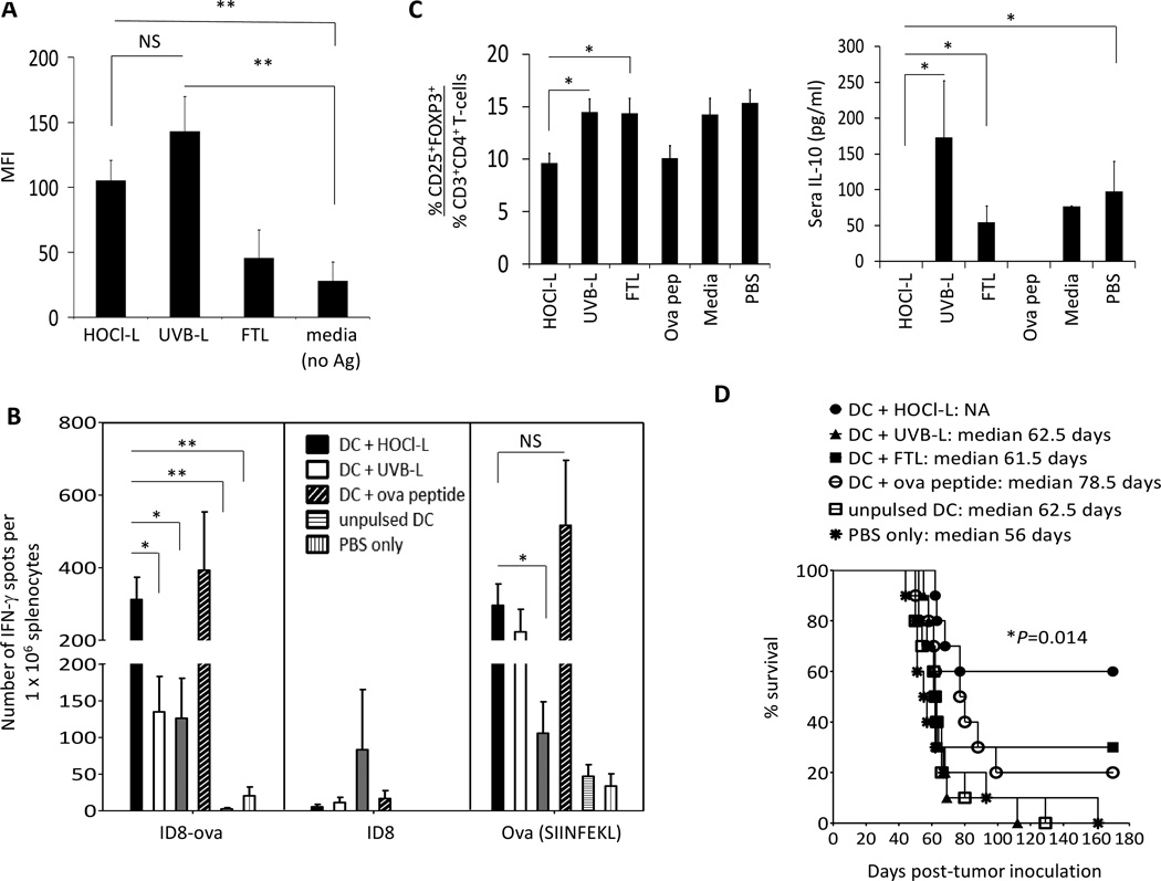
Experimental design: We compared the ability of DCs to engulf three different tumor lysate preparations, produce T-helper 1 (TH1)-priming cytokines and chemokines, stimulate mixed leukocyte reactions (MLR), and finally elicit T-cell responses capable of controlling tumor growth in vivo.

Results: We showed that DCs engulfed HOCl-oxidized lysate most efficiently stimulated robust MLRs, and elicited strong tumor-specific IFN-γ secretions in autologous T cells. These DCs produced the highest levels of TH1-priming cytokines and chemokines, including interleukin (IL)-12. Mice vaccinated with HOCl-oxidized ID8-ova lysate-pulsed DCs developed T-cell responses that effectively controlled tumor growth. Safety, immunogenicity of autologous DCs pulsed with HOCl-oxidized autologous tumor lysate (OCDC vaccine), clinical efficacy, and progression-free survival (PFS) were evaluated in a pilot study of five subjects with recurrent ovarian cancer. OCDC vaccination produced few grade 1 toxicities and elicited potent T-cell responses against known ovarian tumor antigens. Circulating regulatory T cells and serum IL-10 were also reduced. Two subjects experienced durable PFS of 24 months or more after OCDC.

Conclusions: This is the first study showing the potential efficacy of a DC vaccine pulsed with HOCl-oxidized tumor lysate, a novel approach in preparing DC vaccine that is potentially applicable to many cancers.


Conflict of interest statement

Disclosure of Potential Conflicts of Interest

No potential conflicts of interest were disclosed.

Figures

Figure 1
Figure 1
DCs pulsed with HOCl-L efficiently present tumor-associated SIINFEKL, stimulated SIINFEKL-specific CD8+ T-cell and prolonged survival in mice. A, mouse bone marrow-derived DCs were pulsed with HOCl-L, UVB-L or FTL lysate for 20 to 24 hours, matured with LPS and FIN-γ and evaluated for the expression of ovalbumin immunodominant SIINFEKL on cell surface (n per group=6). B, evaluation of the number of IFN-γ secreting tumor-reactive cells in the spleens of mice following DC-lysate vaccinations. Each vaccination group was represented by different color spot and the mean result was indicated as a line in each group. Data was from 2 independent experiments with 5 mice in each group. C, left panel, the % of CD3+CD4+CD25+ FOXP3+ Treg cells was quantified in spleens 2 weeks after the 3rd vaccine. Data was the mean of 3 independent experiments with 5 mice in each experiment. C, right panel, evaluation of IL-10 in sera 2 weeks following 3rd vaccine (n per group=15). D, 3 weeks after live ID8-ova tumor cell inoculation in the peritoneal, mice were vaccinated with different DC-lysate vaccines and their % survival was evaluated. PBS only (star) and unpulsed DC (open square) groups served as controls. Each curve represented data from 2 independent experiments of 5 mice per group per experiment (n per group=10). Data was presented as mean ± standard error of the mean (SEM). Double asterisks indicate results that were highly significant at P<0.001 and single asterisks indicate results that were significant at P<0.05.
Figure 2
Figure 2
HOCl-oxidized tumor lysates enhanced DC uptake, improved cytokine and chemokine productions of DCs, stimulated potent MLRs and elicited tumor-specific FIN-γ secretions from autologous T-cells. A, normal donor DCs were cocultured with HOCl-oxidized SKOV-3 cells [HOCl-L], UVB-irradiated SKOV-3 cells [UVB-L] or freeze-thawed SKOV-3 lysate [FTL] for 4 hours at 37°C or 4°C to determine the % tumor cells/ lysate uptake by DCs. Bar chart was the quantitative representation of the average uptake from 6 different normal individuals. B, cytokine and chemokine profiles of lysate-pulsed DCs from 6 different normal individuals were assessed following 8 hours of LPS and FIN-γ activation. C, stimulation of MLRs by DCs (the averaged of six different normal individuals) pulsed with different tumor lysates preparations or unpulsed mature or immature. D, FIN-γ responses from autologous T-cells after 10 days of coculture with lysate-pulsed DCs or unpulsed DCs (i.e. media, no antigen) to live breast (MDA-231) and ovarian (OVCAR2) tumor lines, and T2 cells pulsed with HER-2/neu369–377 peptide or unpulsed. Data was presented as mean ± SEM. Asterisks indicate results that were significant at P<0.05.
Figure 3
Figure 3
Subject DCs that were pulsed with HOCl-L matured normally with LPS and FIN-γ and exhibited a Th1 cytokine and chemokine profile. DCs prepared for subjects and pulsed with HOCl-oxidized autologous tumor lysate (i.e. OCDC vaccine) in the clinics were thawed and cultured in CellGenix DC media without GM-CSF and IL-4 for 24 hours and evaluated for cytokine and chemokine secretions. Each subject was represented by different color data point.
Figure 4
Figure 4
A, OCDC vaccine schedule of subjects. B, line graph showed regression or stabilization of 6 of 13 tumor metastatic deposits at 128 days (i.e. at EOS II) in subject S4, and CT imaging showed 3 additional nodules with central necrosis/cavitation (enlarged picture) at 128 days. C, taking into account the entire salvage treatment sequence of surgery, adjuvant therapy and ODCD, subject S2 experienced a second progression-free survival (PFS) of 36 versus an initial PFS of 26 months
Figure 5
Figure 5
Tumor-specific T-cells were elicited in subjects following OCDC vaccination. A, subjects’ PBMCs from pre-vaccine and EOS were evaluated for FIN-γ secreting cells in response to autologous DCs pulsed with autologous HOCl-oxidized lysate or to unpulsed autologous DCs. PBMCs alone were used as specificity control. The results were expressed as number of FIN-γ spots per 1×10⁵ human PBLs. B, subjects’ PBMCs taken from pre-vaccine and EOS were assessed for Th1 (FIN-γ), Th2 cytokines (IL-10, IL4 and IL-5) and IL-17. C, ratio of tumor-reactive IFN-g secreting CD3+ cells and Treg cells in subjects’ PBMCs pre- and post-vaccinations. D, measurement of serum IL-10 in subjects at pre-vaccination and EOS.
Figure 6
Figure 6
OCDC vaccines were Th1 priming and primed HER-2/neu specific T-cells that could be expanded ex vivo. A, PBMCs from HLA-A2+ subjects S3 and S4 were further tested for recognition to various HLA-A2 restricted ovarian TAAs. B, five rounds of OCDC vaccinations were able to prime HER-2/neu 369-specific CD8+ T-cells that could further be expanded in vitro following 10 days of stimulation with HER-2/neu369–377 peptide, IL-7 and IL-15 (10ng/ml each). C, expression of various ovarian TAAs was confirmed in the primary tumor of subject S4 with immunohistochemistry. Asterisks indicate results that were significant at P<0.05.

References

    1. Lopez MN, Pereda C, Segal G, Munoz L, Aguilera R, Gonzalez FE, et al. Prolonged survival of dendritic cell-vaccinated melanoma patients correlates with tumor-specific delayed type IV hypersensitivity response and reduction of tumor growth factor beta-expressing T-cells. J Clin Oncol. 2009;27:945–952. - PubMed
    1. Barth RJ, Jr., Fisher DA, Wallace PK, Channon JY, Noelle RJ, Gui J, et al. A randomized trial of ex vivo CD40L activation of a dendritic cell vaccine in colorectal cancer patients: tumor-specific immune responses are associated with improved survival. Clin Cancer Res. 2010;16:5548–5556. - PMC - PubMed
    1. Fadul CE, Fisher JL, Hampton TH, Lallana EC, Li Z, Gui J, et al. Immune response in patients with newly diagnosed glioblastoma multiforme treated with intranodal autologous tumor lysate-dendritic cell vaccination after radiation chemotherapy. J Immunother. 2011;34:382–389. - PMC - PubMed
    1. Hernando JJPT, Kübler K, Offergeld R, Schlebusch H, Bauknecht T. Vaccination with autologous tumour antigen-pulsed dendritic cells in advanced gynaecological malignancies: clinical and immunological evaluation of a phase I trial. Cancer Immunol Immunother. 2002;51:45–52. - PMC - PubMed
    1. Kandalaft LE, Powell DJ, Jr., Chiang CL-L, Tanyi J, Kim S, Bosch M, et al. Autologous Lysate-pulsed Dendritic Cell Vaccination Followed by Adoptive Transfer of Vaccine-primed Ex Vivo Costimulated T-cells in Recurrent Ovarian Cancer. Oncoimmunology. 2013;1(2):e22664. - PMC - PubMed

Publication types

MeSH terms