Review
. 2013 Oct 1;5(10):a010363.
doi: 10.1101/cshperspect.a010363.

Translesion DNA polymerases

Affiliations
Review

Translesion DNA polymerases

Myron F Goodman et al. Cold Spring Harb Perspect Biol. .

Abstract

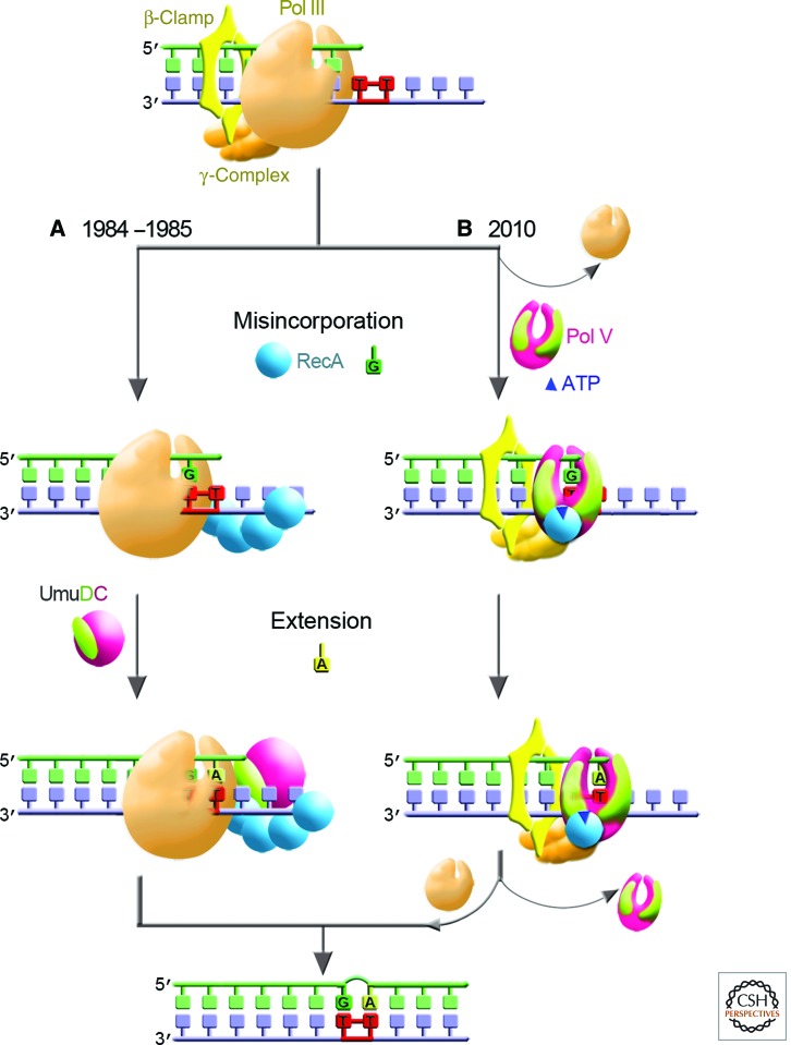
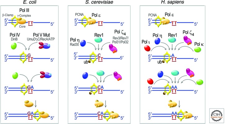
Living cells are continually exposed to DNA-damaging agents that threaten their genomic integrity. Although DNA repair processes rapidly target the damaged DNA for repair, some lesions nevertheless persist and block genome duplication by the cell's replicase. To avoid the deleterious consequence of a stalled replication fork, cells use specialized polymerases to traverse the damage. This process, termed "translesion DNA synthesis" (TLS), affords the cell additional time to repair the damage before the replicase returns to complete genome duplication. In many cases, this damage-tolerance mechanism is error-prone, and cell survival is often associated with an increased risk of mutagenesis and carcinogenesis. Despite being tightly regulated by a variety of transcriptional and posttranslational controls, the low-fidelity TLS polymerases also gain access to undamaged DNA where their inaccurate synthesis may actually be beneficial for genetic diversity and evolutionary fitness.


Figures

Figure 1.
Figure 1.
Two-step model for UV mutagenesis in E. coli. (A) The two-step model for UV mutagenesis proposed in 1984/1985 assumed that TLS in vivo is performed by the replicative Pol III. The first step, nucleotide misincorporation opposite a 3′ T of a CPD, was hypothesized to be mediated by the RecA protein (represented as blue sphere). The misincorporated base was subsequently fixed as a mutation in a second extension/bypass step that was dependent on UmuC (purple) and UmuD (lime) (Bridges and Woodgate 1984, 1985a,b). (B) Subsequent studies revealed that rather than being accessory factors of Pol III, the products of umuDC genes encode a bona fide DNA polymerase, Pol V (shown as purple UmuC and two lime UmuD′ subunits assembled in the shape of a right hand) (Reuven et al. 1999; Tang et al. 1999), which interacts with RecA* in trans (not shown) from which molecules of RecA (blue sphere) and ATP (dark blue triangle) are transferred from the 3′-filament tip to generate Pol V Mut (Jiang et al. 2009). Pol V Mut can perform both the (mis)insertion and extension steps of TLS. After traversing the damaged DNA, Pol V Mut is replaced by Pol III holenzyme, which resumes high-fidelity chromosomal duplication.
Figure 2.
Figure 2.
Evolutionarily conserved roles of TLS polymerases. Replicative polymerases such as E. coli Pol III or eukaryotic Pol ε stall at the site of a DNA lesion. TLS polymerases are recruited to the site via interactions with the replicative sliding processivity clamp (β-subunit in E. coli and PCNA in eukaryotes). E. coli has a choice of three TLS polymerases: Pol II, Pol IV, and Pol V Mut (composed of UmuD′2C-RecA-ATP) (Jiang et al. 2009). S. cerevisiae also has a choice of three TLS polymerases: Pol ζ4, Rev1, and Pol η. In humans, at least five TLS polymerases can be recruited to sites of arrested replication, including Pol η, Pol ι, Pol κ, Rev1, and Pol ζ4. Additional human DNA polymerases, such as Pol β, Pol λ, Pol θ, and/or Pol ν (Maga et al. 2007; Seki and Wood 2008; Shtygasheva et al. 2008; Yamanaka et al. 2010; Hogg et al. 2011; Villani et al. 2011), may also facilitate TLS under certain conditions, but these have been omitted for clarity. The likelihood that this insertion step is error-prone, as shown for E. coli and S. cerevisiae, or error-free, as shown for humans, will depend on the DNA lesion encountered and the polymerase used for TLS. The extension step may be facilitated by the same enzyme that performed the (mis)insertion or by a different polymerase. Once the nascent DNA chain has been extended beyond the lesion, the TLS polymerase is replaced by the cell’s replicative DNA polymerase so as to complete genome duplication.
Figure 3.
Figure 3.
Structural insights into TLS polymerases and their mutagenic specificity. In panels A and B, the main domains are color-coded: (red) palm; (green) thumb; (blue) fingers; (purple) little finger. (A) Crystal structure of human Pol η in a ternary complex with a CPD. In this view, the 3′T of the CPD is in the active site and is correctly paired with incoming dATP (PDB: 3MR3) (Biertümpfel et al. 2010). (Rust) The template strand; (olive green) the primer; (yellow) the incoming dNTP. (Burgundy stick) The position of the CPD; (small blue spheres) the metal ions. The protein backbone is represented by the ribbon surrounded by the semitransparent solvent-accessible surface. (B) Crystal structure of the S. solfataricus Dpo4 in a ternary complex with DNA containing a benzo[a]pyrene lesion (brown) and incoming nucleotide (yellow) (PDB: 1S0M_BP-2) (Ling et al. 2004b). As can be seen, the benzo[a]pyrene lesion is flipped into the major groove, so as to accommodate base pairing. (C) Human Pol ι making a G:T mispair (PDB:3GV8); note that the template dT and incoming dG are both in an anti conformation (Kirouac and Ling 2009) and the mispair is stabilized through hydrogen bonds with Gln59. (D) Arg357-directed dC incorporation by human Rev1 (PDB: 3GQC) (Swan et al. 2009). (C,D) (White/gray) The protein surface; (dotted lines) hydrogen bonds.
Figure 4.
Figure 4.
Domain organization of selected TLS polymerases. (A) Prokaryotes; (B) archaea; (C) eukaryotes. The name and phylogenetic family relationship of each of the TLS polymerases along with number of amino acid residues in each polymerase are indicated on the right-hand side of the figure. The structural catalytic domains present in all of the polymerases are color-coded as follows: (red) palm; (green) thumb; (blue) finger; (purple) little finger; (violet) 3′–5′ exonuclease of Pol II; (yellow) amino-terminal domain of Pol II and Rev3; (pink) uncharacterized catalytic domain of Rev3. Additional domains involved in localization and regulation of the TLS polymerases are as follows: (teal octagon) breast cancer-associated protein-1 carboxy-terminal domain (BRCT); (gray rectangle) nuclear localization signal (NLS); (green diamond) ubiquitin binding motif (UBM); (red diamond) ubiquitin binding zinc-finger motif (UBZ); (grayish oval) zinc finger (ZF); (gold oval) PCNA/β-clamp motif; (brown or blue rhomboid) Rev7 binding region; (olive rhomboid) Rev1-interacting region. Species: Ec, E. coli; Ss, S. solfataricus; Sc, S. cerevisiae; Hs, H. sapiens.

References

    1. Anderson WF, Prince DB, Yu H, McEntee K, Goodman MF 1994. Crystallization of DNA polymerase II from Escherichia coli. J Mol Biol 238: 120–122 - PubMed
    1. Baranovskiy AG, Lada AG, Siebler HM, Zhang Y, Pavlov YI, Tahirov TH 2012. DNA polymerase δ and ζ switch by sharing accessory subunits of DNA polymerase δ. J Biol Chem 287: 17281–17287 - PMC - PubMed
    1. Beaber JW, Hochhut B, Waldor MK 2004. SOS response promotes horizontal dissemination of antibiotic resistance genes. Nature 427: 72–74 - PubMed
    1. Bebenek K, Kunkel TA 2004. Functions of DNA polymerases. Adv Protein Chem 69: 137–165 - PubMed
    1. Becherel OJ, Fuchs RP, Wagner J 2002. Pivotal role of the β-clamp in translesion DNA synthesis and mutagenesis in E. coli cells. DNA Repair 1: 703–708 - PubMed

Publication types

Substances