

Reduced subcortical glutamate/glutamine in adults with autism spectrum disorders: a [¹H]MRS study
- PMID: 23838890
- PMCID: PMC3731785
- DOI: 10.1038/tp.2013.53
Reduced subcortical glutamate/glutamine in adults with autism spectrum disorders: a [¹H]MRS study
Erratum in
-
Reduced subcortical glutamate/glutamine in adults with autism spectrum disorders: a [(1)H]MRS study.Transl Psychiatry. 2014 Feb 18;4(2):e364. doi: 10.1038/tp.2014.7. Transl Psychiatry. 2014. PMID: 24548879 Free PMC article. No abstract available.
Abstract
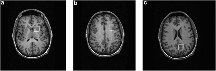
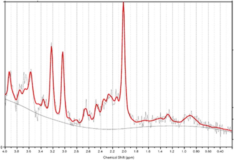
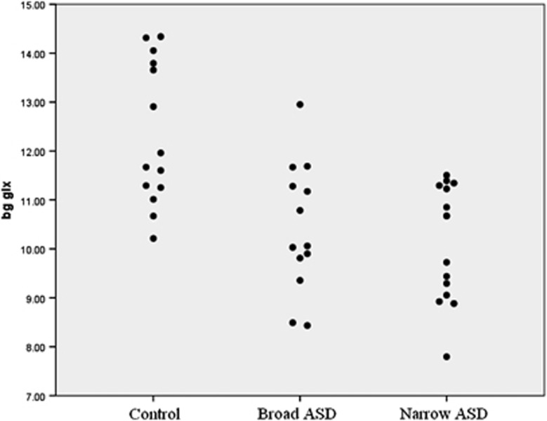
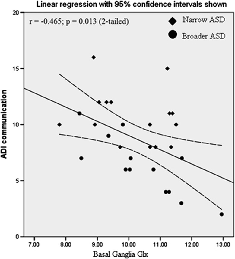
Dysfunctional glutamatergic neurotransmission has been implicated in autism spectrum disorder (ASD). However, relatively few studies have directly measured brain glutamate in ASD adults, or related variation in glutamate to clinical phenotype. We therefore set out to investigate brain glutamate levels in adults with an ASD, comparing these to healthy controls and also comparing results between individuals at different points on the spectrum of symptom severity. We recruited 28 adults with ASD and 14 matched healthy controls. Of those with ASD, 15 fulfilled the 'narrowly' defined criteria for typical autism, whereas 13 met the 'broader phenotype'. We measured the concentration of the combined glutamate and glutamine signal (Glx), and other important metabolites, using proton magnetic resonance spectroscopy in two brain regions implicated in ASD--the basal ganglia (including the head of caudate and the anterior putamen) and the dorsolateral prefrontal cortex--as well as in a parietal cortex 'control' region. Individuals with ASD had a significant decrease (P<0.001) in concentration of Glx in the basal ganglia, and this was true in both the 'narrow' and 'broader' phenotype. Also, within the ASD sample, reduced basal ganglia Glx was significantly correlated with increased impairment in social communication (P=0.013). In addition, there was a significant reduction in the concentration of other metabolites such as choline, creatine (Cr) and N-acetylaspartate (NAA) in the basal ganglia. In the dorsolateral prefrontal cortex, Cr and NAA were reduced (P<0.05), although Glx was not. There were no detectable differences in Glx, or any other metabolite, in the parietal lobe control region. There were no significant between-group differences in age, gender, IQ, voxel composition or data quality. In conclusion, individuals across the spectrum of ASD have regionally specific abnormalities in subcortical glutamatergic neurotransmission that are associated with variation in social development.
Figures
References
-
- World Health Organization ICD-10 International Statistical Classification of Diseases and Related Health Problems10th ednWHO: Geneva, Switzerland; 1993
-
- Baron-Cohen S, Scott FJ, Allison C, Williams J, Bolton P, Matthews FE, et al. Prevalence of autism-spectrum conditions: UK school-based population study. Br J Psychiatry. 2009;194:500–509. - PubMed
-
- Hallahan B, Daly EM, McAlonan G, Loth E, Toal F, O'Brien F, et al. Brain morphometry volume in autistic spectrum disorder: a magnetic resonance imaging study of adults. Psychol Med. 2009;39:337–346. - PubMed
-
- Barttfeld P, Wicker B, Cukier S, Navarta S, Lew S, Sigman M. A big-world network in ASD: dynamical connectivity analysis reflects a deficit in long-range connections and an excess of short-range connections. Neuropsychologia. 2011;49:254–263. - PubMed
Publication types
MeSH terms
Substances
Grants and funding
LinkOut - more resources
Full Text Sources
Other Literature Sources
Miscellaneous
