Comment
. 2013 Jul-Sep;7(3):173-83.
doi: 10.4161/fly.25241. Epub 2013 Jul 9.

Steroids as external temporal codes act via microRNAs and cooperate with cytokines in differential neurogenesis

Affiliations
Comment

Steroids as external temporal codes act via microRNAs and cooperate with cytokines in differential neurogenesis

Mariya M Kucherenko et al. Fly (Austin). 2013 Jul-Sep.

Abstract

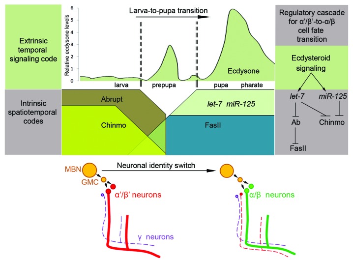
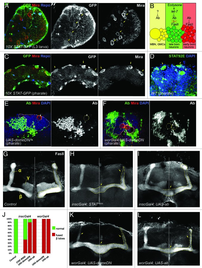
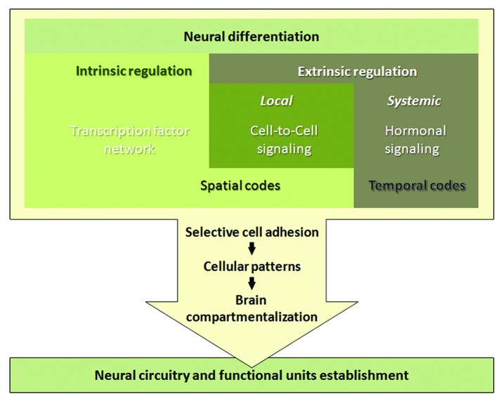
The generation of neuronal cell diversity is controlled by interdependent mechanisms, including cell intrinsic programs and environmental cues. During development, the astonishing variety of neurons is originated according to a precise timetable that is managed by a complex network of genes specifying individual types of neurons. Different neurons express specific sets of transcription factors, and they can be recognized by morphological characteristics and spatial localization, but, most importantly, they connect to each other and form functional units in a stereotyped fashion. This connectivity depends, mostly, on selective cell adhesion that is strictly regulated. While intrinsic factors specifying neuronal temporal identity have been extensively studied, an extrinsic temporal factor controlling neuronal temporal identity switch has not been shown. Our data demonstrate that pulses of steroid hormone act as a temporal cue to fine-tune neuronal cell differentiation. Here we also provide evidence that extrinsic JAK/STAT cytokine signaling acts as a spatial code in the process. Particularly, in Drosophila mushroom bodies, neuronal identity transition is controlled by steroid-dependent microRNAs that regulate spatially distributed cytokine-dependent signaling factors that in turn modulate cell adhesion. A new era of neuronal plasticity assessment via managing external temporal cues such as hormones and cytokines that specify individual types of neurons might open new possibilities for brain regenerative therapeutics.

Keywords: Drosophila mushroom body; JAK/STAT cytokine signaling; differential cell adhesion; microRNA let-7; steroid hormone ecdysone; temporal identity switch.


Figures

None
Figure 1. Model of differential neurogenesis regulation by cooperation of developmentally controlled temporal systemic signaling and intrinsic spatiotemporal codes. Scheme represents the chronologically regulated signaling cascade controlling α′/β′ to α/β neuronal identity switch in the Drosophila MB that takes place at the larva-to-pupa developmental transition. Amount of ecdysone at different stages of development is represented as relative levels (scheme adopted from ref. 15). Developmentally regulated pulse of the steroid hormone ecdysone acts as an extrinsic temporal signaling code to activate expression of miRNAs from the let-7 complex in the differentiating MB neurons.- Temporally induced miRNAs let-7 and miR-125 are intrinsic spatiotemporal codes that downregulate at least two BTB domain containing transcription factors Abrupt and Chinmo,, which allows for the α′/β′ to α/β neuronal cell fate transition. Cell adhesion molecule FasII is downstream of let-7/Abrupt signaling. During larval stages Abrupt suppresses FasII expression allowing for early-born lobes to be formed, while at the pupal stage downregulation of Abrupt allows FasII expression and promote α/β neuronal differentiation.
None
Figure 2. JAK/STAT signaling is involved in Abrupt regulation during MB development. (A, C and D) In Drosophila brain JAK/STAT signaling activity [marked with 10xSTAT-GFP reporter in (A) and (C)] and STAT92E antibody staining (D) is detected at larval and pharate stages. JAK/STAT signaling is active in neuroblasts [marked with anti-Miranda (red) and glial cells marked with anti-Repo (blue) in (A) and (C) or determined based on morphology and nuclear DAPI staining in (D)]. Yellow arrows indicate both JAK/STAT signaling activity and neuroblast location. (B) Schematic drawing of Abrupt expression pattern and its regulation by previously described regulatory factors in the MB neuronal body cluster. Abrupt expression is restricted to the early born γ, α′/β′ MB (red colored) neurons where it functions as a negative regulator of FasII (cell adhesion molecule) expression. At the larva-to-pupa transition developmentally regulated ecdysteroid signaling induces expression of miRNA let-7 in the α/β (green colored) neurons. let-7 negatively regulates Abrupt which allows for FasII expression, necessary for MB neurons to undergo cell fate transition into α/β. The question mark depicted on the scheme inquires whether spatially distributed cytokine signaling acts in the concert with temporally regulated hormonal stimuli to adjust Abrupt activity in the mushroom body neuroblast (MBN) and ganglion mother cells (GMCs) (yellow). (E and F) anti-Abrupt staining (green) is elevated in the MBN upon JAK/STAT signaling downregulation achieved by overexpression of dominant negative form of dome (F) in comparison to the control (E). Circles show MBN location [marked also with anti-Miranda (red)], arrows point to anti-Abrupt staining inside the MBN, white dashed line outlines Ab-negative area in the MB cell body clusters [note smaller area in (F) in comparison to (E)]. (G-L) Both, downregulation of JAK/STAT signaling (H, J and K) and overexpression of the transcription factor Abrupt in the neuroblasts (I, J and L) causes similar morphological defects detected with anti-FasII staining in the adult brains in comparison to control [UAS-domeDN/TM6 in (G)]. White vertical line indicates position of the midline, dashed yellow line shows α/β MB lobes, yellow arrows point to slim α/β and fused β MB lobes.
None
Figure 3. Model of spatiotemporal regulation of differential neurogenesis. Differentiation of the neural stem cell progenitor into a specific neuron subtype depends on concerted action of intrinsic and extrinsic programs. Intrinsic regulation is achieved via combination of multiple transcription factors that are hierarchically specified during organismal development starting from establishing the anterior-posterior and dorsal-ventral polarity that creates gradients of morphogens and induces expression of gap, pair-rule and Hox genes, and subsequently assembling a set of differentially expressed transcription factors, combination of which produces the unique code for a certain neuronal subtype. This code is additionally adjusted by extrinsic cell-to-cell signaling, for example Notch for binary cell fate decision or JAK/STAT cytokine signaling for neuronal cell type specification. This unique code constantly changes in response to internal and external conditions that coordinate the development of the whole organism. Hormones are great temporal code candidates, as they direct all major developmental steps. The combination of spatial and temporal codes in neuronal precursors allows certain types of neurons to be born at exact place and time, which is critical for brain morphogenesis. For normal brain function, these neurons must cluster and synapse in a stereotyped fashion, which predominantly depends on selective cell adhesion. As a result of establishment of brain compartments and differential neuronal connections, functional neural circuits are created that process all kinds of information and control behavior, learning, memory and plasticity of each individual.

Comment on

References

    1. Leone DP, Srinivasan K, Chen B, Alcamo E, McConnell SK. The determination of projection neuron identity in the developing cerebral cortex. Curr Opin Neurobiol. 2008;18:28–35. doi: 10.1016/j.conb.2008.05.006. - DOI - PMC - PubMed
    1. Passante L, Gaspard N, Degraeve M, Frisén J, Kullander K, De Maertelaer V, et al. Temporal regulation of ephrin/Eph signalling is required for the spatial patterning of the mammalian striatum. Development. 2008;135:3281–90. doi: 10.1242/dev.024778. - DOI - PubMed
    1. Lin S, Lee T. Generating neuronal diversity in the Drosophila central nervous system. Dev Dyn. 2012;241:57–68. doi: 10.1002/dvdy.22739. - DOI - PubMed
    1. Franco SJ, Müller U. Shaping our minds: stem and progenitor cell diversity in the mammalian neocortex. Neuron. 2013;77:19–34. doi: 10.1016/j.neuron.2012.12.022. - DOI - PMC - PubMed
    1. Hoshino M. Neuronal subtype specification in the cerebellum and dorsal hindbrain. Dev Growth Differ. 2012;54:317–26. doi: 10.1111/j.1440-169X.2012.01330.x. - DOI - PubMed

Publication types

MeSH terms

LinkOut - more resources