Review
. 2013 Aug;14(8):529-41.
doi: 10.1038/nrm3619. Epub 2013 Jul 10.

Mending broken hearts: cardiac development as a basis for adult heart regeneration and repair

Affiliations
Review

Mending broken hearts: cardiac development as a basis for adult heart regeneration and repair

Mei Xin et al. Nat Rev Mol Cell Biol. 2013 Aug.

Abstract

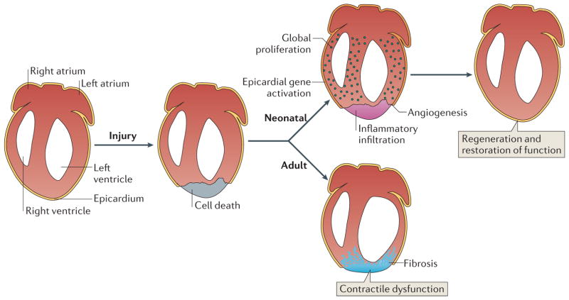
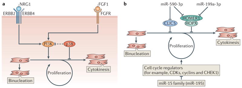
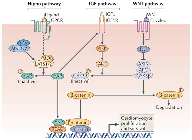
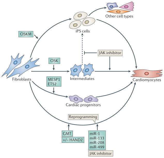
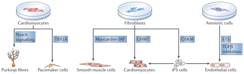
As the adult mammalian heart has limited potential for regeneration and repair, the loss of cardiomyocytes during injury and disease can result in heart failure and death. The cellular processes and regulatory mechanisms involved in heart growth and development can be exploited to repair the injured adult heart through 'reawakening' pathways that are active during embryogenesis. Heart function has been restored in rodents by reprogramming non-myocytes into cardiomyocytes, by expressing transcription factors (GATA4, HAND2, myocyte-specific enhancer factor 2C (MEF2C) and T-box 5 (TBX5)) and microRNAs (miR-1, miR-133, miR-208 and miR-499) that control cardiomyocyte identity. Stimulating cardiomyocyte dedifferentiation and proliferation by activating mitotic signalling pathways involved in embryonic heart growth represents a complementary approach for heart regeneration and repair. Recent advances in understanding the mechanistic basis of heart development offer exciting opportunities for effective therapies for heart failure.


Conflict of interest statement

Competing interests statement

The authors declare no competing financial interests.

Figures

Figure 1
Figure 1. Regeneration of the mammalian neonatal heart
The neonatal mouse heart possesses regenerative capacity. In neonatal mice, the early response to cardiac injury-induced cardiomyocyte loss includes inflammatory infiltration, activation of epicardial-specific genes and angiogenesis. Global proliferation of cardiomyocytes in the injured heart replaces the scar tissue with cardiomyocytes and restores cardiac function within 3 weeks after the injury. The regenerative capacity of the mammalian neonatal heart declines with age, whereas cardiac fibrosis increases with age in response to injury. The regenerative potential of the mouse heart is lost when injury occurs 7 days postnatally or later during adulthood, when fibrotic scar tissue replaces the lost cardiomyocytes and leads to reduced cardiac function.
Figure 2
Figure 2. Regulation of cardiomyocyte proliferation
a | Regulation of cardiomyocyte proliferation by fibroblast growth factor 1 (FGF1) and neuregulin 1 (NRG1). Inhibition of the MAPK p38 in the presence of FGF1 or the activation of NGR1 signalling promotes cardiomyocyte re-entry into the cell cycle by activating PI3K, leading to DNA synthesis and cytokinesis. However, most cardiomyocytes in mice become binucleated shortly after birth as a consequence of DNA replication without cell division. b | Regulation of cardiomyocyte proliferation by microRNAs (miRNAs). miRNAs can positively (miR-590-3p and miR-199a-3p) or negatively (miR-15 family, including miR-195) regulate cardiomyocyte proliferation. miR-590-3p and miR-199a-3p promote cardiomyocyte proliferation by inhibiting the expression of genes encoding proteins that inhibit cell proliferation such as HOMER1, HOP homeobox (HOPX) and chloride intracellular channel 5 (CLIC5). The miR-15 family of miRNAs inhibits the cell cycle, and thus cardiomyocyte proliferation, by downregulating genes encoding proteins that activate the cell cycle. CDKs, cyclin-dependent kinases; CHEK1, checkpoint kinase 1; FGFR, FGF receptor.
Figure 3
Figure 3. Regulation of cardiomyocyte proliferation by the Hippo pathway
Activation of the Hippo pathway, which includes the scaffold protein Salvador (SAV) and the kinases mammalian STE20-like protein kinase 1 (MST1), MST2, large tumour suppressor 1 (LATS1) and LATS2, results in the phosphorylation and nuclear exclusion of the transcriptional co-activator Yes-associated protein (YAP) to impede cardiomyocyte proliferation. Deletion of the upstream scaffold protein SAV or the inhibitory Hippo kinases LATS2, MST1 and MST2 promotes cardiomyocyte proliferation, as unphosphorylated YAP can activate the expression of genes in the WNT signalling pathway by interacting with β-catenin. Genetic deletion of YAP in the embryonic heart inhibits cardiomyocyte proliferation, leading to myocardial hypoplasia and embryonic lethality. Overexpression of YAP also promotes cardiomyocyte proliferation through the activation of pro-growth signalling pathways such as those driven by WNT and insulin-like growth factor 1 (IGF1). APC, adenomatosis polyposis coli; AXIN, axis inhibition protein; GPCR, G protein-coupled receptor; DVL, Dishevelled; GSK3β; glycogen synthase kinase 3 β; IGF1R, IGF1 receptor; LEF, lymphoid enhancer-binding factor; MOB, Mps one binder; TCF, T cell factor. Dashed icons represent components of the complex that phosphorylates β-catenin to promote its degradation, and dashed lines indicate that additional factors are involved.
Figure 4
Figure 4. Reprogramming fibroblasts into cardiomyocytes
Several approaches have been used to reprogramme non-myocytes, such as adult fibroblasts and mouse embryonic fibroblasts (MEFs), into cardiomyocytes. In one method, the four pluripotency genes, OCT4, SOX2, Kruppel-like factor 4 (KLF4) and MYC (collectively referred to as OSKM factors), are overexpressed to reprogramme fibroblasts into induced pluripotent stem (iPS) cells, which subsequently differentiate into cardiomyocytes. A modification to this protocol is to overexpress OCT4, SOX2 and KLF4 (OSK factors), but not the oncoprotein MYC, to reduce the potential of oncogenic transformation. Transient overexpression of OSK factors epigenetically converts MEFs into plastic intermediates, without the cells becoming iPS cells. A Janus kinase (JAK) inhibitor helps prevent the formation of iPS cells from these intermediates (indicated by the dashed line) and promotes their differentiation towards cardiomyocytes. Alternatively, the forced expression of the basic helix loop helix (bHLH) transcription factor mesoderm posterior 1 (MESP1) and the transcription factor ETS2 in human dermal fibroblasts can induce their differentiation into beating cardiomyocytes via cardiac progenitors. Another approach involves the direct reprogramming of fibroblasts into cardiomyocytes, using transcription factors that specify the cardiac lineage and the differentiation of cardiomyocytes during embryogenesis. Forced expression of GATA4, myocyte-specific enhancer 2C (MEF2C) and T-box 5 (TBX5) with HAND2 (GHMT) or without HAND2 (GMT) can convert adult cardiac and skin fibroblasts into beating cardiomyocyte-like cells. GHMT or GMT factors can reprogramme non-cardiac cells into functional cardiomyocytes in vivo and improve cardiac function in response to injury. Cardiomyocytes have also been reprogrammed by introducing the microRNAs (miRNAs) miR-1, miR-133, miR-208 and miR-499 into cardiac fibroblasts. The addition of a JAK inhibitor enhanced reprogramming by these miRNAs.
Figure 5
Figure 5. Reprogramming different cardiac cell types
Progress has been made in using transcriptional reprogramming to generate different specialized cell types that are essential for restoring cardiac function in an injured heart. For the conduction system, pacemaker cells and Purkinje fibres can be generated by reprogramming. Overexpression of the transcription factor T-box 18 (TBX18) directly reprogrammes rodent ventricular cardiomyocytes, in vitro and in vivo, into cells resembling pacemaker cells from the sinoatrial node, without passage of cells through a pluripotent state. The activation of Notch signalling in newborn rodent cardiomyocytes,in vitro and in vivo, confers a Purkinje cell-like phenotype. For the vasculature, smooth muscle cells and endothelial cells can be produced from fibroblasts by reprogramming. Smooth muscle cells can be generated by forced expression of the cardiovascular-specific co-activator myocardin. Myocardin activates smooth muscle-specific genes through association with the transcription factor serum response factor (SRF). Adult fibroblasts can also be reprogrammed into an incomplete induced pluripotent state by transient forced overexpression of the four induced pluripotent stem (iPS) cell factors OCT4, SOX2, Kruppel-like factor 4 (KLF4) and MYC (OSKM factors); endothelial cells then form under permissive culture conditions. Endothelial cells can also be generated from amniotic cells by forced overexpression of ETS transcription factors, in combination with the inhibition of transforming growth factor-β (TGFβ) signalling, without cells transiting through an iPS cell state. Finally, cardiomyocytes can be generated by reprogramming fibroblasts indirectly via an iPS cell state that is induced by OSKM factors, or directly with the cardiac-specific transcription factors GATA4, HAND2, myocyte-specific enhancer factor 2C (MEF2C) and T-box 5 (TBX5) (collectively referred to as GHMT) (see also FIG. 4).

References

    1. Lopez AD, Mathers CD, Ezzati M, Jamison DT, Murray CJ. Global and regional burden of disease and risk factors, 2001: systematic analysis of population health data. Lancet. 2006;367:1747–1757. - PubMed
    1. Hill JA, Olson EN. Cardiac plasticity. N Engl J Med. 2008;358:1370–1380. - PubMed
    1. Segers VF, Lee RT. Stem-cell therapy for cardiac disease. Nature. 2008;451:937–942. - PubMed
    1. Laflamme MA, Murry CE. Heart regeneration. Nature. 2011;473:326–335. - PMC - PubMed
    1. Wollert KC, Drexler H. Cell therapy for the treatment of coronary heart disease: a critical appraisal. Nature Rev Cardiol. 2010;7:204–215. - PubMed

Publication types