. 2013 Aug 30;288(35):25477-25489.
doi: 10.1074/jbc.M113.454850. Epub 2013 Jul 9.

Intrinsic selectivity of Notch 1 for Delta-like 4 over Delta-like 1

Affiliations

Intrinsic selectivity of Notch 1 for Delta-like 4 over Delta-like 1

Marie Blanke Andrawes et al. J Biol Chem. .

Abstract

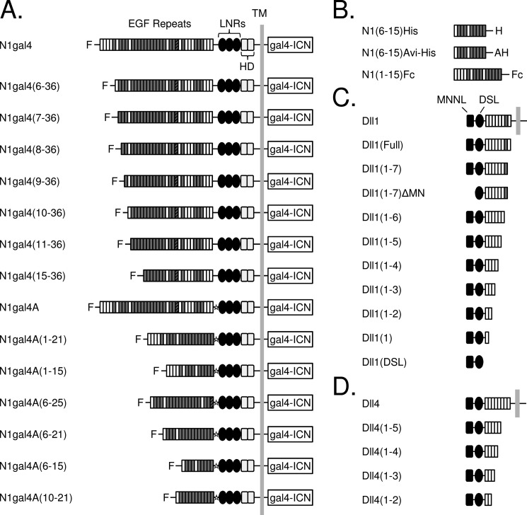
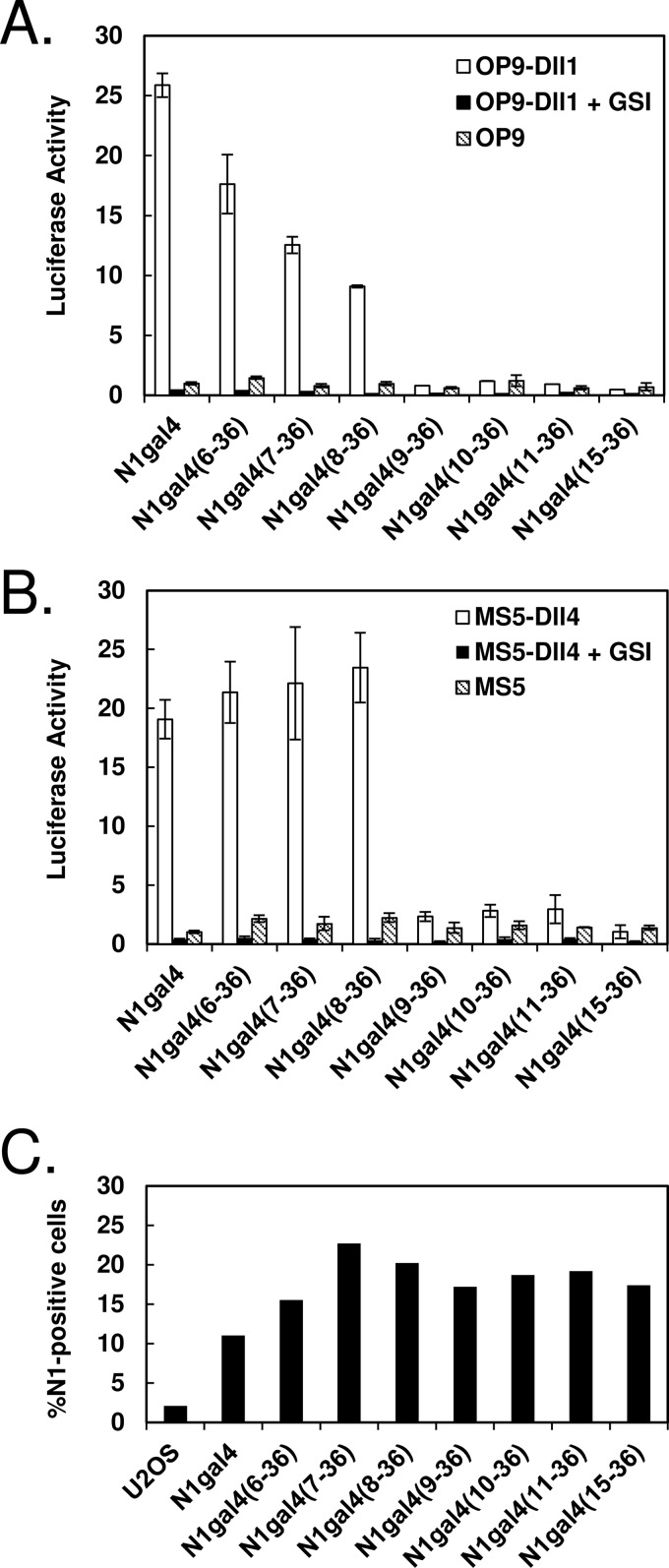
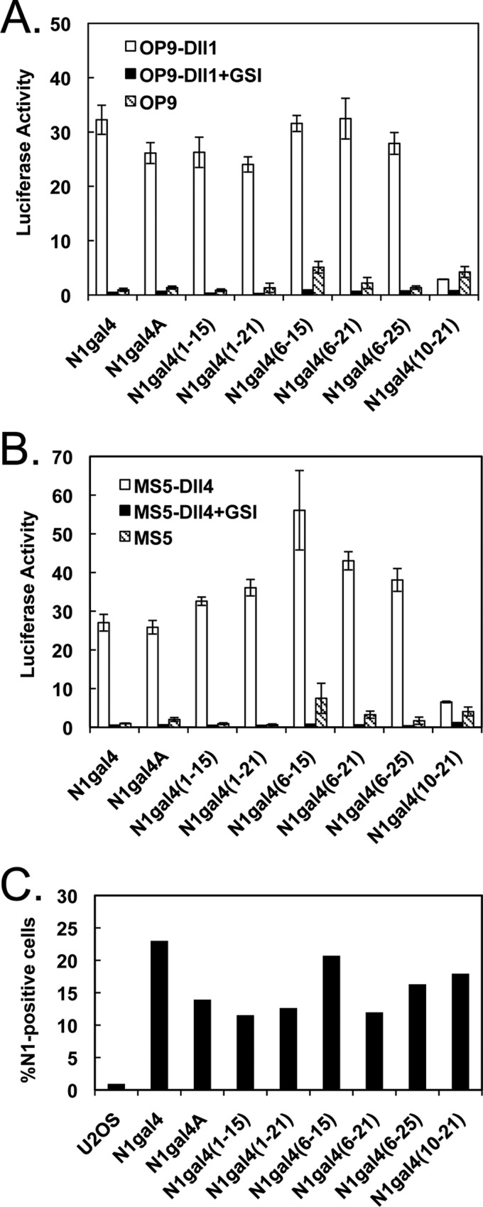
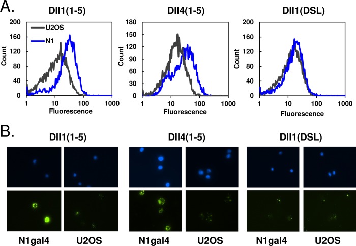
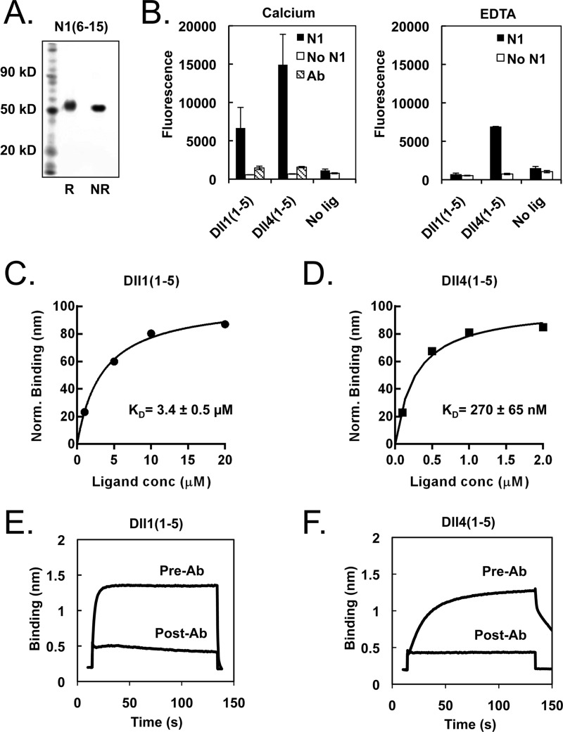
Notch signaling makes critical contributions to cell fate determination in all metazoan organisms, yet remarkably little is known about the binding affinity of the four mammalian Notch receptors for their three Delta-like and two Jagged family ligands. Here, we utilized signaling assays and biochemical studies of purified recombinant ligand and receptor molecules to investigate the differences in signaling behavior and intrinsic affinity between Notch1-Dll1 and Notch1-Dll4 complexes. Systematic deletion mutagenesis of the human Notch1 ectodomain revealed that epidermal growth factor (EGF) repeats 6-15 are sufficient to maintain signaling in a reporter assay at levels comparable with the full-length receptor, and identified important contributions from EGF repeats 8-10 in conveying an activating signal in response to either Dll1 or Dll4. Truncation studies of the Dll1 and Dll4 ectodomains showed that the MNNL-EGF3 region was both necessary and sufficient for full activation. Plate-based and cell binding assays revealed a specific, calcium-dependent interaction between cell-surface and recombinant Notch receptors and ligand molecules. Finally, direct measurement of the binding affinity of Notch1 EGF repeats 6-15 for Dll1 and Dll4 revealed that Dll4 binds with at least an order of magnitude higher affinity than Dll1. Together, these studies give new insights into the features of ligand recognition by Notch1, and highlight how intrinsic differences in the biochemical behavior of receptor-ligand complexes can influence receptor-mediated responses of developmental signaling pathways.

Keywords: Biolayer Interferometry; Cell Signaling; Cell Surface Receptor; Cell-Cell Interaction; Notch Pathway; Receptor Structure-Function; Receptor-Ligand Interaction; Recombinant Protein Expression.


Figures

FIGURE 1.
FIGURE 1.
Domain organization of ligand and receptor constructs. A, Notch molecules used in signaling assays. The N1gal4 construct was created by fusing a gal4 promoter to the intracellular region of Notch1 (gal4-ICN), and EGF repeats were systematically truncated from the N terminus. Repeats containing a consensus calcium-binding motif are shaded in gray, and EGF25, which contains an extra cysteine residue, is indicated with a striped pattern. “F” indicates the presence of an N-terminal FLAG epitope. C-terminal truncations were constructed using a Notch1-gal4A variant, which contains a restriction site (indicated with a star) between the EGF repeat region and the NRR. B, Notch molecules used in binding assays. The Notch extracellular truncation spanning EGF repeats 6–15, inclusive, was designed for use in protein-protein binding experiments, along with a N1(1–15)Fc fusion protein (R&D Biosystems). C and D, ligand molecules used in binding and signaling assays. C, human Delta-like 1 was systematically truncated from the C terminus, along with a variant lacking the N-terminal MNNL domain. D, C-terminal truncations of human Delta-like 4. LNRs, LIN12-Notch repeats; HD, heterodimerization domain.
FIGURE 2.
FIGURE 2.
Signaling of N-terminal Notch truncations. A and B, ligand-induced activation of N-terminal N1gal4 truncations. U2OS cells were transiently transfected with receptor variants, co-cultured with ligand-expressing or control cells, and the resulting activation was measured via luciferase activity. A, Notch N-terminal truncations activated with OP9-Dll1 cells. Gradual loss of activity can be seen with removal of EGF repeats 6 and 7, and complete loss of activity with removal of EGF8. B, Notch N-terminal truncations activated with MS5-Dll4 cells. Activity equivalent to full-length N1gal4 is observed upon deletion of EGF repeats 6 and 7, but deletion of EGF8 abolishes signaling. All results are shown normalized to co-culture with non-ligand-bearing OP9 or MS5 cells, and treatment with the γ-secretase inhibitor compound E (GSI, 1 μm) inhibits activation in all receptor constructs tested. C, surface expression of truncated receptors. Surface expression was detected via flow cytometry, using an antibody to the Notch1 NRR (WC-75) and a FITC-conjugated secondary antibody. Percentage of Notch1-positive cells is indicated on the y axis.
FIGURE 3.
FIGURE 3.
Signaling of C-terminal Notch truncations. A and B, ligand-induced activation of C-terminal N1gal4 truncations. Luciferase assays were performed as described in the legend to Fig. 2. A, Notch C-terminal truncations activated with OP9-Dll1 cells. Activity comparable with full-length N1gal4A is observed in C-terminal truncations up to EGF15, and truncating from both ends resulted in the identification of the EGF repeat 6–15 fragment, the smallest Notch1 fragment identified with activity comparable with the full-length ectodomain. B, Notch C-terminal truncations activated with MS5-Dll4 cells. The observed activation pattern is similar to OP9-Dll1 results. All data are shown normalized to co-culture with non-ligand-bearing OP9 or MS5 cells, and treatment with 1 μm GSI inhibits activation in all receptor constructs tested. C, surface expression of truncated receptors. Surface expression was detected via flow cytometry, using an antibody to the Notch1 NRR (WC-75) and a FITC-conjugated secondary antibody. Percentage of Notch1-positive cells is indicated on the y axis.
FIGURE 4.
FIGURE 4.
Signaling of ligand truncations. A, non-reducing SDS-PAGE gel of purified Dll1 proteins. B, non-reducing SDS-PAGE gel of purified Dll4 proteins. Multiple bands are present as a result of N-linked glycosylation, which was not observed in Dll1. C, signaling induced by Dll1 truncations. Purified Dll1 proteins were plated at an equivalent molar concentration and used to activate N1gal4 U2OS stable cells. Resulting luciferase activity is shown on the y axis, normalized to activation with plated human IgG. C-terminal truncation up to EGF3 had little effect on signaling, whereas removal of the N-terminal MNNL domain (ΔMN) abolished activity. D, signaling induced by Dll4 truncations, performed as in C. C-terminal truncations up to EGF3 also maintained full signaling capacity, whereas removal of EGF3 abolished signaling. E and F, concentration dependence of ligand-induced signaling. Dll1(1–5) and Dll4(1–5) were plated at varying concentrations and incubated with U2OS cells transfected with either full-length N1gal4 or N1gal4(6–15). The resulting luciferase activity is shown as a function of concentration, fitted with a one-site exponential binding model. Calculated EC50 values for N1gal4 were 227 ± 175 nm (Dll1) and 47 ± 22 nm (Dll4); for N1gal4(6–15), 281 ± 82 nm (Dll1) and 47 ± 33 nm (Dll4).
FIGURE 5.
FIGURE 5.
Ligand binding to Notch cells. A, detection of purified ligands binding to Notch-bearing cells. Biotinylated ligands were coupled to yellow fluorescent avidin beads and incubated with Notch1-gal4 U2OS or U2OS control cells. When analyzed via flow cytometry, the N1gal4 cells incubated with Dll1(1–5) and Dll4(1–5) showed a shift in fluorescence relative to U2OS control cells, which was not observed with the shorter Dll1(DSL) ligand. B, visualization of ligand-cell binding with fluorescence microscopy. Top and bottom rows show DAPI and FITC staining, respectively. Beads coated with Dll1(1–5) or Dll4(1–5) both bound preferentially to Notch-bearing cells over control cells, whereas Dll1(DSL)-coated beads did not.
FIGURE 6.
FIGURE 6.
Measurement of Notch-ligand interactions using purified proteins. A, SDS-PAGE gel of purified N1(6–15) protein under reducing and non-reducing conditions. B, ELISA binding assay. Functional ELISAs were performed on plated N1(6–15), bound with biotinylated Dll1(1–5) or Dll4(1–5) preclustered with Neutravidin-HRP. The resulting interaction is calcium-dependent and inhibited by blocking antibody. C and D, quantification of Notch-ligand binding affinity via biolayer interferometry (BLI). Biotinylated N1(6–15) was immobilized on a streptavidin biosensor and bound to varying concentrations of Dll1(1–5) or Dll4(1–5). The resulting concentration-response plots were fitted with a one-site exponential binding model and normalized for comparison, yielding KD values of 270 ± 65 nm for N1-Dll4 and 3.4 ± 0.5 μm for N1-Dll1 interactions. E and F, specificity of observed Notch-ligand binding. Treatment with the ligand-blocking antibody WC-613 (1 μg/ml) inhibited binding of Dll1 (5 μm) or Dll4 (2 μm) to N1(6–15).

References

    1. Artavanis-Tsakonas S., Rand M. D., Lake R. J. (1999) Notch signaling. Cell fate control and signal integration in development. Science 284, 770–776 - PubMed
    1. Bray S. J. (2006) Notch signalling. A simple pathway becomes complex. Nat. Rev. Mol. Cell Biol. 7, 678–689 - PubMed
    1. Aster J. C., Pear W. S., Blacklow S. C. (2008) Notch signaling in leukemia. Annu. Rev. Pathol. 3, 587–613 - PMC - PubMed
    1. Penton A. L., Leonard L. D., Spinner N. B. (2012) Notch signaling in human development and disease. Semin. Cell Dev. Biol. 23, 450–457 - PMC - PubMed
    1. Fukuda D., Aikawa E., Swirski F. K., Novobrantseva T. I., Kotelianski V., Gorgun C. Z., Chudnovskiy A., Yamazaki H., Croce K., Weissleder R., Aster J. C., Hotamisligil G. S., Yagita H., Aikawa M. (2012) Notch ligand Delta-like 4 blockade attenuates atherosclerosis and metabolic disorders. Proc. Natl. Acad. Sci. U.S.A. 109, E1868–E1877 - PMC - PubMed

Publication types

MeSH terms

LinkOut - more resources