

Design, synthesis, and application of the trimethoprim-based chemical tag for live-cell imaging
- PMID: 23839994
- PMCID: PMC3800088
- DOI: 10.1002/9780470559277.ch130019
Design, synthesis, and application of the trimethoprim-based chemical tag for live-cell imaging
Abstract
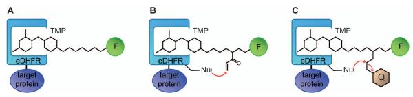
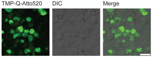
Over the past decade, chemical tags have been developed to complement the use of fluorescent proteins in live-cell imaging. Chemical tags retain the specificity of protein labeling achieved with fluorescent proteins through genetic encoding, but provide smaller, more robust tags and modular use of organic fluorophores with high photon output and tailored functionalities. The trimethoprim-based chemical tag (TMP-tag) was initially developed based on the high affinity interaction between E. coli dihydrofolate reductase and the antibiotic trimethoprim and was subsequently rendered covalent and fluorogenic via proximity-induced protein labeling reactions. To date, the TMP-tag is one of the few chemical tags that enable intracellular protein labeling and high-resolution live-cell imaging. Here we describe the general design, chemical synthesis, and application of TMP-tag for live-cell imaging. Alternate protocols for synthesizing and using the covalent and the fluorogenic TMP-tags are also included.
Keywords: chemical tag; fluorescence microscopy; live‐cell imaging; protein label.
© 2013 by John Wiley & Sons, Inc.
Figures
Similar articles
-
A fluorogenic TMP-tag for high signal-to-background intracellular live cell imaging.ACS Chem Biol. 2013 Aug 16;8(8):1704-12. doi: 10.1021/cb300657r. Epub 2013 Jun 19. ACS Chem Biol. 2013. PMID: 23745575 Free PMC article.
-
The covalent trimethoprim chemical tag facilitates single molecule imaging with organic fluorophores.Biophys J. 2014 Jan 7;106(1):272-8. doi: 10.1016/j.bpj.2013.11.4488. Biophys J. 2014. PMID: 24411259 Free PMC article.
-
Second-generation covalent TMP-tag for live cell imaging.J Am Chem Soc. 2012 Aug 22;134(33):13692-9. doi: 10.1021/ja303374p. Epub 2012 Aug 9. J Am Chem Soc. 2012. PMID: 22873118 Free PMC article.
-
Exploiting Covalent Chemical Labeling with Self-Labeling Proteins.Annu Rev Biochem. 2025 Jun;94(1):29-58. doi: 10.1146/annurev-biochem-030222-121016. Epub 2025 Mar 19. Annu Rev Biochem. 2025. PMID: 40106680 Review.
-
Chemical biology-based approaches on fluorescent labeling of proteins in live cells.Mol Biosyst. 2013 May;9(5):862-72. doi: 10.1039/c2mb25422k. Mol Biosyst. 2013. PMID: 23318293 Review.
Cited by
-
On-Demand Targeting: Investigating Biology with Proximity-Directed Chemistry.J Am Chem Soc. 2016 Mar 23;138(11):3610-22. doi: 10.1021/jacs.5b12608. Epub 2016 Mar 7. J Am Chem Soc. 2016. PMID: 26907082 Free PMC article.
-
Fluorescent Trimethoprim Conjugate Probes To Assess Drug Accumulation in Wild Type and Mutant Escherichia coli.ACS Infect Dis. 2016 Oct 14;2(10):688-701. doi: 10.1021/acsinfecdis.6b00080. Epub 2016 Aug 16. ACS Infect Dis. 2016. PMID: 27737551 Free PMC article.
References
-
- Ando T, Tsukiji S, Tanaka T, Nagamune T. Construction of a small-molecule-integrated semisynthetic split intein for in vivo protein ligation. Chem Commun (Camb): 2007;4995:4997. - PubMed
-
- Baccanari DP, Daluge S, King RW. Inhibition of dihydrofolate reductase: Effect of reduced nicotinamide adenine dinucleotide phosphate on the selectivity and affinity of diaminobenzylpyrimidines. Biochemistry. 1982;21:5068–5075. - PubMed
-
- Bloch KD. Digestion of DNA with restriction endonucleases. Curr Protoc Immunol. 2001 Chapter 10: Unit 10 18. - PubMed
-
- Calloway NT, Choob M, Sanz A, Sheetz MP, Miller LW, Cornish VW. Optimized fluorescent trimethoprim derivatives for in vivo protein labeling. Chembiochem. 2007;8:767–774. - PubMed
Publication types
MeSH terms
Substances
Grants and funding
LinkOut - more resources
Full Text Sources
Other Literature Sources
Medical
Research Materials
Miscellaneous
