. 2013 Jul 23;110(30):12301-6.
doi: 10.1073/pnas.1309947110. Epub 2013 Jul 9.

Validated near-atomic resolution structure of bacteriophage epsilon15 derived from cryo-EM and modeling

Affiliations

Validated near-atomic resolution structure of bacteriophage epsilon15 derived from cryo-EM and modeling

Matthew L Baker et al. Proc Natl Acad Sci U S A. .

Abstract

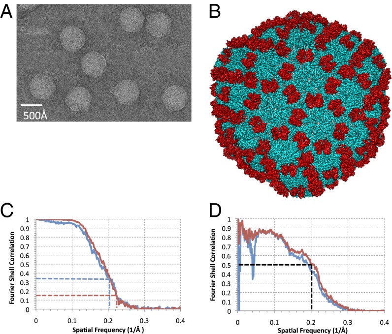
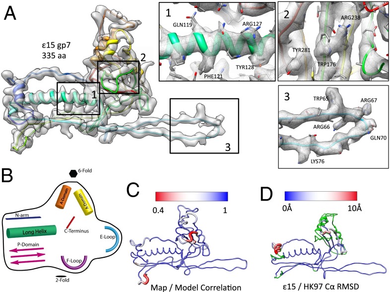
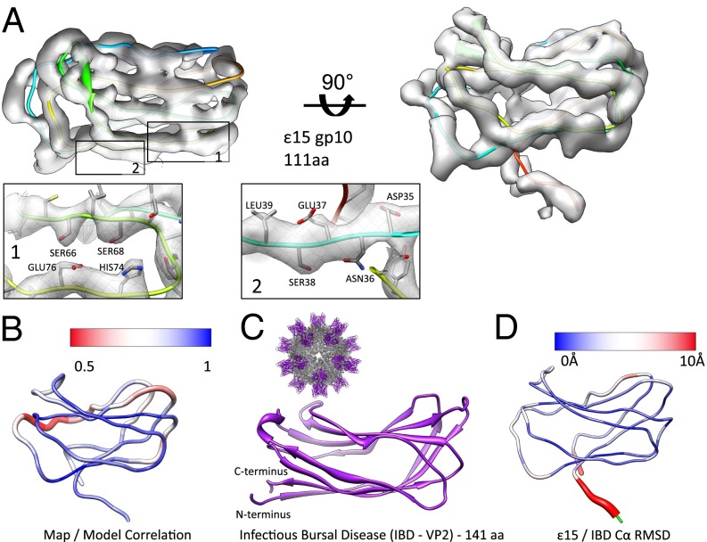
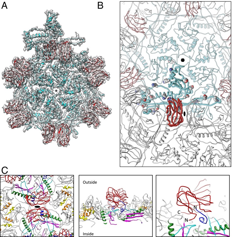
High-resolution structures of viruses have made important contributions to modern structural biology. Bacteriophages, the most diverse and abundant organisms on earth, replicate and infect all bacteria and archaea, making them excellent potential alternatives to antibiotics and therapies for multidrug-resistant bacteria. Here, we improved upon our previous electron cryomicroscopy structure of Salmonella bacteriophage epsilon15, achieving a resolution sufficient to determine the tertiary structures of both gp7 and gp10 protein subunits that form the T = 7 icosahedral lattice. This study utilizes recently established best practice for near-atomic to high-resolution (3-5 Å) electron cryomicroscopy data evaluation. The resolution and reliability of the density map were cross-validated by multiple reconstructions from truly independent data sets, whereas the models of the individual protein subunits were validated adopting the best practices from X-ray crystallography. Some sidechain densities are clearly resolved and show the subunit-subunit interactions within and across the capsomeres that are required to stabilize the virus. The presence of the canonical phage and jellyroll viral protein folds, gp7 and gp10, respectively, in the same virus suggests that epsilon15 may have emerged more recently relative to other bacteriophages.

Keywords: EMAN; PHENIX; Pathwalker; gold standard; validation.


Conflict of interest statement

The authors declare no conflict of interest.

Figures

Fig. 1.
Fig. 1.
Structure of ε15. (A) A typical image of ε15 300 kV at 50,000× magnification from the JEM3200FSC electron cryomicroscope. (B) The complete reconstruction of ε15 reveals the capsid protein gp7 (cyan) and the staple protein gp10 (red). (C) The FSC between the two independent datasets used for our current reconstruction is shown in red. The FSC between the previous ε15 density map (EMDB ID code 5003) and our current ε15 density map (EMDB ID code 5678) is shown in blue. The dashed lines correspond to the gold standard criterion for resolution estimate. (D) A comparison of density simulated at 3.5 Å resolution from our all-atom model versus our current density map is shown (red). Application of an inner mask removes the low-resolution dip, which can be attributed to the DNA (blue).
Fig. 2.
Fig. 2.
Structure of the capsid protein, gp7. (A) An averaged gp7 density map is superimposed on the gp7 (Chain C) molecular model (rainbow representation, N–C terminus). Insets (–3) show zoomed-in views of the typical regions with the unaveraged density and corresponding model of one gp7 subunit (Chain C). (B) A cartoon diagram illustrates the structural domains of gp7 shared with other tailed dsDNA bacteriophage (Fig. S7). (C) Correlation values were computed between the map and model on a per amino acid level, then averaged for all seven subunits in the asymmetric unit. Regions shown in blue had a strong correlation between the map and model, as opposed to regions in red, which had a weak correlation between the map and model. (D) A gp7 model is colored based on the Cα rmsd between the capsid proteins from ε15 and HK97. Variations range from low to high rmsd (blue to red); green regions of the model were not used in computing the rmsd.
Fig. 3.
Fig. 3.
Structure of the staple protein, gp10. (A) An averaged gp10 density map is shown in two views with the corresponding model (Chain C in rainbow representation). Individual sections of the model are highlighted (insets 1 and 2). (B) Correlation values were computed between the map and model on a per amino acid basis, then averaged for all seven subunits in the asymmetric unit. Regions shown in blue had a strong correlation between the map and model, as opposed to the red regions, which had a weak correlation between the map and model. (C) VP2 of infectious bursal disease virus (PDB ID code 2DF7) shows a similar jellyroll fold with four strands on the top and bottom. A full view of the infections bursal disease virus is shown (Inset, Upper Left). (D) The gp10 model is colored based on Cα rmsd between gp10 and the corresponding domain of 2DF7. The largest differences are shown in red. Residues highlighted in green were not used in computing the rmsd.
Fig. 4.
Fig. 4.
ε15 subunit interactions. (A) An asymmetric unit for ε15 consists of seven gp7 (cyan) and seven gp10 (red) subunits. For illustration purposes, additional gp10 subunits are highlighted to illustrate the dimeric relationship. (B) One gp10 (bold red subunit) and one gp7 (bold cyan subunit) are shown with potential salt bridges shown as spheres (Table S2). (C) The gp10 dimer is highlighted in red and spans the twofold axis (Left). Four gp7 subunits are directly below a single gp10. The N-arm regions of gp7 are colored in dark blue and flank the outer strands of gp7. The color annotations for gp7 are the same as defined in Fig. 2B. Directly below the gp10 dimers are the gp7 E-loops (cyan). Rotating 90° shows a slice through the capsid with both gp10 termini positioned at the twofold interface, protruding down into the gp7 subunits (Center). A zoomed-in view of this region highlights the positioning of the N-arm of gp7 and the termini of gp10 (Right).

References

    1. Wommack KE, Colwell RR. Virioplankton: Viruses in aquatic ecosystems. Microbiol Mol Biol Rev. 2000;64(1):69–114. - PMC - PubMed
    1. Hendrix RW, Duda RL. Bacteriophage HK97 head assembly: A protein ballet. Adv Virus Res. 1998;50:235–288. - PubMed
    1. Jiang W, et al. Structure of epsilon15 bacteriophage reveals genome organization and DNA packaging/injection apparatus. Nature. 2006;439(7076):612–616. - PMC - PubMed
    1. Jiang W, et al. Backbone structure of the infectious epsilon15 virus capsid revealed by electron cryomicroscopy. Nature. 2008;451(7182):1130–1134. - PubMed
    1. Wikoff WR, et al. Topologically linked protein rings in the bacteriophage HK97 capsid. Science. 2000;289(5487):2129–2133. - PubMed

Publication types

Associated data

LinkOut - more resources