

Early-stage primary signet ring cell carcinoma of the colon
- PMID: 23840131
- PMCID: PMC3699035
- DOI: 10.3748/wjg.v19.i24.3895
Early-stage primary signet ring cell carcinoma of the colon
Abstract
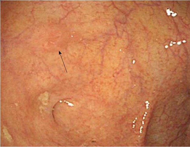
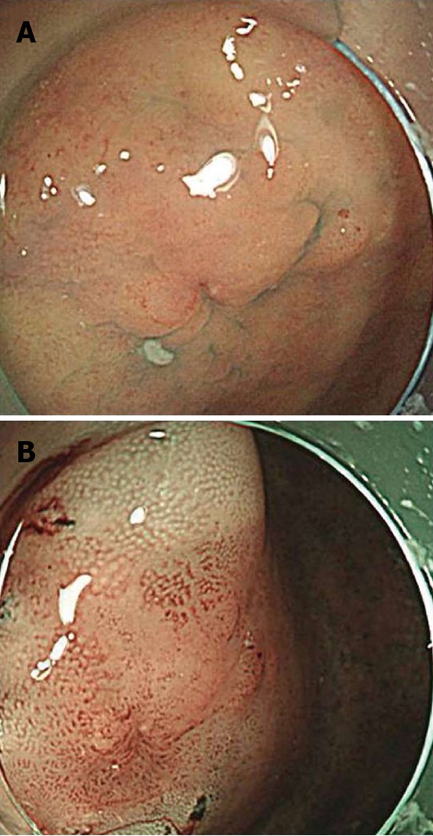
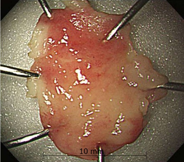
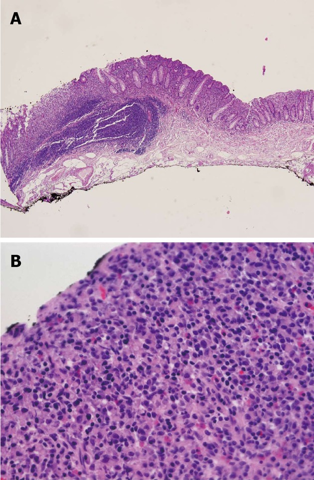
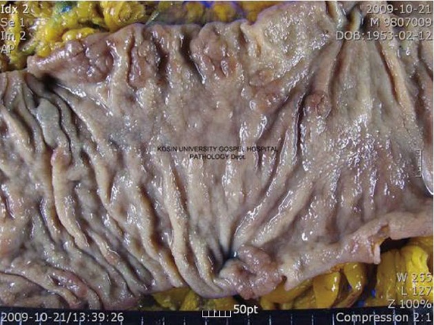
Primary signet ring cell carcinoma of the colorectum detected at an early stage is very rare; most cases are detected at an advanced stage. Therefore, its prognosis is poorer than that of ordinary colorectal cancer. A 56-year-old Korean man was seen at this hospital for management of signet ring cell carcinoma of the colon. Colonoscopic examination revealed a IIa-like, ill-defined and flatly elevated 9-mm residual tumor in the cecum. Endoscopic mucosal resection was preformed. Pathological examination of the resected specimen revealed signet ring cell carcinoma that had invaded the lamina propria without venous or perineural invasion. Abdominal computed tomography (CT) and positron CT showed no evidence of primary lesions or distant metastasis. An additional laparoscopic right-hemicolectomy was performed; no residual tumor or lymph node metastasis was found. We report a case of primary signet ring cell carcinoma of the colon detected at an early stage and provide a review of the literature.
Keywords: Colon carcinoma; Early stage; Endoscopic mucosal resection; Primary carcinoma; Signet ring cell carcinoma.
Figures
Similar articles
-
Primary signet-ring cell carcinoma of the colon at early stage: a case report and a review of the literature.World J Gastroenterol. 2006 Jun 7;12(21):3446-9. doi: 10.3748/wjg.v12.i21.3446. World J Gastroenterol. 2006. PMID: 16733868 Free PMC article. Review.
-
Early-stage primary signet ring cell carcinoma of the colon with magnifying endoscopic findings.Gastrointest Endosc. 2019 Sep;90(3):529-531. doi: 10.1016/j.gie.2019.05.010. Epub 2019 May 18. Gastrointest Endosc. 2019. PMID: 31108090 No abstract available.
-
Early stage signet ring cell carcinoma of the colon examined by magnifying endoscopy with narrow-band imaging: a case report.BMC Gastroenterol. 2015 Jul 24;15:86. doi: 10.1186/s12876-015-0317-z. BMC Gastroenterol. 2015. PMID: 26205810 Free PMC article.
-
A Case of Primary Colonic Signet Ring Cell Carcinoma in a Young Man which Preoperatively Mimicked Phlebosclerotic Colitis.Acta Med Okayama. 2019 Aug;73(4):361-365. doi: 10.18926/AMO/56939. Acta Med Okayama. 2019. PMID: 31439960
-
Signet-ring cell carcinoma of the colon 7mm in size with peritonitis carcinomatosa.J Gastroenterol. 2002;37(7):550-5. doi: 10.1007/s005350200085. J Gastroenterol. 2002. PMID: 12162414 Review.
Cited by
-
Three-Dimensional CT Texture Analysis to Differentiate Colorectal Signet-Ring Cell Carcinoma and Adenocarcinoma.Cancer Manag Res. 2019 Dec 13;11:10445-10453. doi: 10.2147/CMAR.S233595. eCollection 2019. Cancer Manag Res. 2019. PMID: 31997883 Free PMC article.
-
Metastatic signet ring cell carcinoma of unknown primary source.BMJ Case Rep. 2014 Feb 17;2014:bcr2013203407. doi: 10.1136/bcr-2013-203407. BMJ Case Rep. 2014. PMID: 24536055 Free PMC article.
-
Prognosis of Signet Ring Cell Carcinoma of the Colon and Rectum and their Distinction of Mucinous Adenocarcinoma with Signet Ring Cells. A Comparative Study.Pathol Oncol Res. 2018 Jul;24(3):609-616. doi: 10.1007/s12253-017-0283-6. Epub 2017 Aug 7. Pathol Oncol Res. 2018. PMID: 28785968
-
Outcomes of Definitive Treatment of Signet Ring Cell Carcinoma of the Rectum: Is Minimal Invasive Surgery Detrimental in Signet Ring Rectal Cancers?Indian J Surg Oncol. 2020 Dec;11(4):597-603. doi: 10.1007/s13193-020-01142-2. Epub 2020 Aug 3. Indian J Surg Oncol. 2020. PMID: 33299278 Free PMC article.
-
Can a Solitary Juvenile Polyp Be Regarded as a Nonmalignant Polyp?ACG Case Rep J. 2022 Dec 26;9(12):e00936. doi: 10.14309/crj.0000000000000936. eCollection 2022 Dec. ACG Case Rep J. 2022. PMID: 36600791 Free PMC article.
References
-
- Anthony T, George R, Rodriguez-Bigas M, Petrelli NJ. Primary signet-ring cell carcinoma of the colon and rectum. Ann Surg Oncol. 1996;3:344–348. - PubMed
-
- Psathakis D, Schiedeck TH, Krug F, Oevermann E, Kujath P, Bruch HP. Ordinary colorectal adenocarcinoma vs. primary colorectal signet-ring cell carcinoma: study matched for age, gender, grade, and stage. Dis Colon Rectum. 1999;42:1618–1625. - PubMed
-
- Chen JS, Hsieh PS, Chiang JM, Yeh CY, Tsai WS, Tang R, Changchien CR, Wu RC. Clinical outcome of signet ring cell carcinoma and mucinous adenocarcinoma of the colon. Chang Gung Med J. 2010;33:51–57. - PubMed
-
- Tung SY, Wu CS, Chen PC. Primary signet ring cell carcinoma of colorectum: an age- and sex-matched controlled study. Am J Gastroenterol. 1996;91:2195–2199. - PubMed
-
- Laufman H, Saphir O. Primary linitis plastica type of carcinoma of the colon. AMA Arch Surg. 1951;62:79–91. - PubMed
Publication types
MeSH terms
LinkOut - more resources
Full Text Sources
Other Literature Sources
