

Epstein-Barr virus negative primary hepatic leiomyoma: case report and literature review
- PMID: 23840159
- PMCID: PMC3703201
- DOI: 10.3748/wjg.v19.i25.4094
Epstein-Barr virus negative primary hepatic leiomyoma: case report and literature review
Abstract
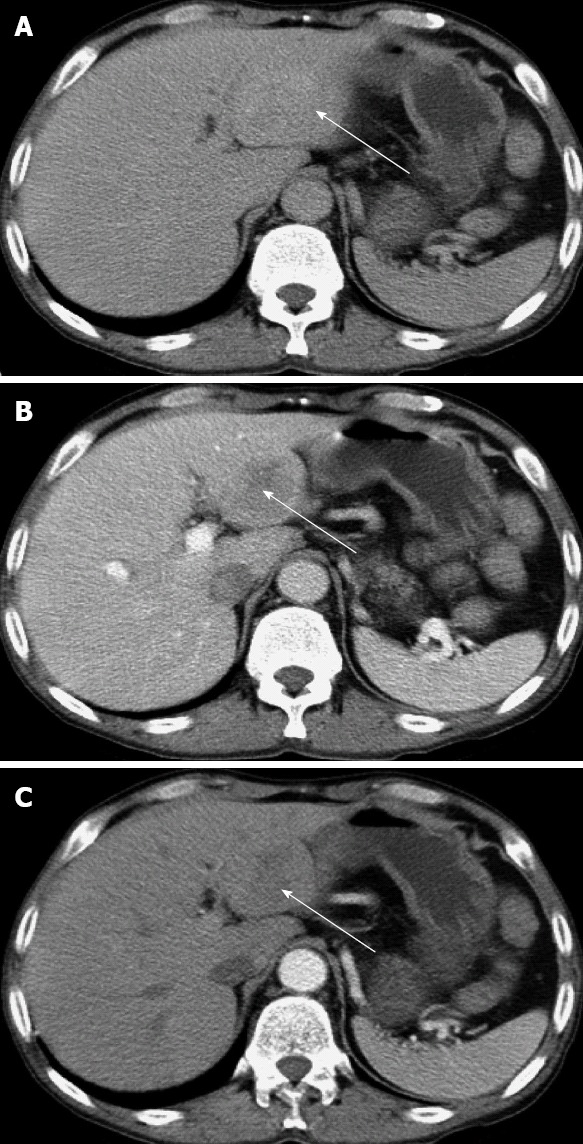
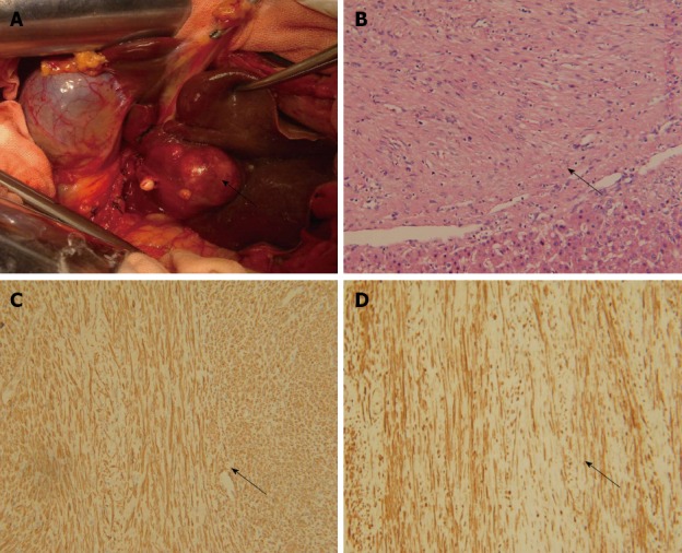
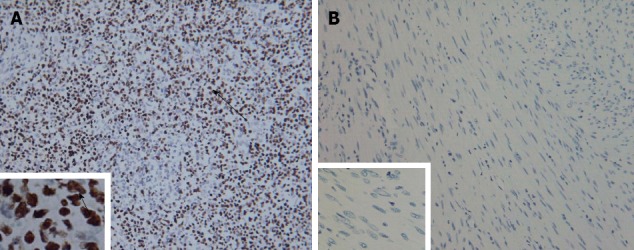
Primary hepatic leiomyoma is a neoplasm of mesenchymal origin and occurs only rarely. Secondary to benign smooth muscle proliferation, it is usually found in adult women and is associated with Epstein-Barr virus (EBV) infection. Here, we report the 29(th) case of primary hepatic leiomyoma with its unique features related to diagnosis, treatment and developmental biology. A 48-year-old man, with an immunocompromised status, complained of pain in the upper quadrant of the abdomen. Serological analysis indicated no presence of hepatitis virus, no human immunodeficiency virus, and no EBV infection. The levels of α-fetoprotein and carcinoembryonic antigen were normal. A mass was detected in segment III of the hepatic lobe by ultrasonography and an abdominal computed tomography scan. Endoscopy had negative findings. Exploratory laparotomy found no existing extrahepatic tumor and left lateral lobectomy was performed. Pathological examination showed the mass to be a typical leiomyoma. The cells were positive for α-smooth muscle actin and desmin, and negative for the makers of gastrointestinal stromal tumor (GIST), including CD117, CD34 and DOG1 (discovered on GIST1). In situ hybridization revealed negative status for EBV-encoded small RNA. After left lateral lobectomy, the patient was not given chemotherapy or radiotherapy. During a 2-year follow-up, no sign of local recurrence or distant metastasis was observed. In conclusion, we report a rare case of primary hepatic leiomyoma in a male patient without EBV infection. Hepatic resection was curative. This case presents data to expand our knowledge concerning the complex and heterogeneous nature of primary liver leiomyoma, indicating that EBV infection is important but neither necessary nor sufficient for the development of primary liver leiomyoma.
Keywords: Cancer diagnosis; Developmental biology; Epstein-Barr virus; Primary hepatic leiomyoma; Tumor resection.
Figures
Similar articles
-
Intracranial Leiomyoma Associated with Epstein-Barr Virus: A Cerebellopontine Angle Mass Presenting with Trigeminal Neuralgia.World Neurosurg. 2020 Sep;141:284-290. doi: 10.1016/j.wneu.2020.05.157. Epub 2020 May 23. World Neurosurg. 2020. PMID: 32450307
-
[Primary leiomyoma of the liver: a case report].Nihon Geka Gakkai Zasshi. 2005 Nov;106(11):725-9. Nihon Geka Gakkai Zasshi. 2005. PMID: 16304825 Japanese.
-
Detection of Epstein Barr virus in an hepatic leiomyomatous neoplasm in an adult human immunodeficiency virus 1-infected patient.Virchows Arch. 1994;425(3):321-5. doi: 10.1007/BF00196156. Virchows Arch. 1994. PMID: 7812519
-
Primary Intraventricular Leiomyoma in an Immunocompetent Patient: First Case Report and Review of the Literature.World Neurosurg. 2016 Jun;90:698.e13-698.e18. doi: 10.1016/j.wneu.2016.01.088. Epub 2016 Feb 5. World Neurosurg. 2016. PMID: 26855311 Review.
-
Primary iris leiomyoma.Surv Ophthalmol. 2017 May-Jun;62(3):366-370. doi: 10.1016/j.survophthal.2016.11.007. Epub 2016 Nov 24. Surv Ophthalmol. 2017. PMID: 27890619 Review.
Cited by
-
Asymptomatic Hepatic Leiomyoma in an Immunocompetent Middle-Aged Woman.ACG Case Rep J. 2022 Nov 24;9(11):e00899. doi: 10.14309/crj.0000000000000899. eCollection 2022 Nov. ACG Case Rep J. 2022. PMID: 36447767 Free PMC article.
-
Primary hepatic leiomyoma in a Chinese female patient without underlying disease: a case report.BMC Surg. 2019 Oct 7;19(1):140. doi: 10.1186/s12893-019-0598-1. BMC Surg. 2019. PMID: 31590641 Free PMC article.
-
Primary hepatic leiomyoma: unusual cause of an intrahepatic mass.Ann Transl Med. 2015 Apr;3(5):73. doi: 10.3978/j.issn.2305-5839.2015.03.40. Ann Transl Med. 2015. PMID: 25992372 Free PMC article.
-
Primary leiomyoma of the liver: a review of a rare tumour.HPB Surg. 2014;2014:959202. doi: 10.1155/2014/959202. Epub 2014 Nov 19. HPB Surg. 2014. PMID: 25505821 Free PMC article. Review.
-
Primary leiomyoma of the liver in an immunocompetent patient.Intractable Rare Dis Res. 2020 Nov;9(4):251-255. doi: 10.5582/irdr.2020.03075. Intractable Rare Dis Res. 2020. PMID: 33139985 Free PMC article.
References
-
- Demel R. Ein operierter fall von leber-myom. Virchows Arch. 1926;261:881–884. doi: 10.1007/BF01892215. - DOI
-
- Imasato M, Tono T, Kano T, Kimura Y, Iwazawa T, Ohnishi T, Nakano Y, Yano H, Okamoto S, Monden T. Primary leiomyoma of the liver: a case report. Nihon Geka Gakkai Zasshi. 2005;106:725–729. - PubMed
-
- Perini MV, Fink MA, Yeo DA, Carvalho CA, Morais CF, Jones RM, Christophi C. Primary liver leiomyoma: a review of this unusual tumour. ANZ J Surg. 2013;83:230–233. - PubMed
Publication types
MeSH terms
LinkOut - more resources
Full Text Sources
Other Literature Sources
Medical
Molecular Biology Databases
