. 2013 Jul 3:4:170.
doi: 10.3389/fimmu.2013.00170. eCollection 2013.

Monitoring the Dynamics of T Cell Clonal Diversity Using Recombinant Peptide:MHC Technology

Affiliations

Monitoring the Dynamics of T Cell Clonal Diversity Using Recombinant Peptide:MHC Technology

J Lori Blanchfield et al. Front Immunol. .

Abstract

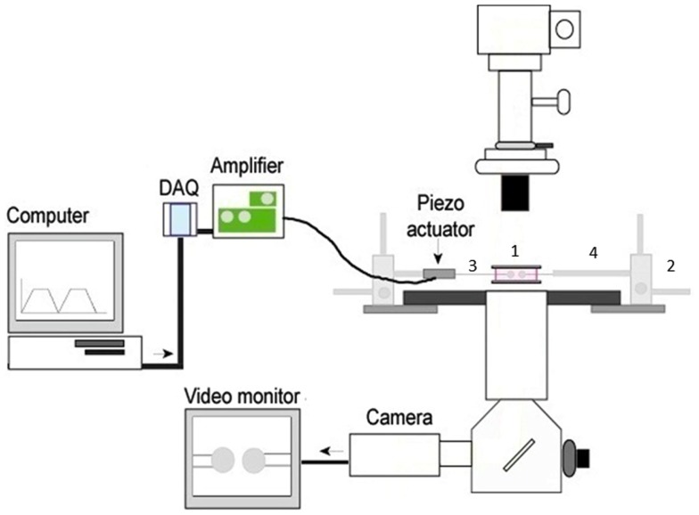
The capacity to probe antigen specific T cells within the polyclonal repertoire has been revolutionized by the advent of recombinant peptide:MHC (pMHC) technology. Monomers and multimers of pMHC molecules can enrich for and identify antigen specific T cells to elucidate the contributions of T cell frequency, localization, and T cell receptor (TCR) affinity during immune responses. Two-dimensional (2D) measurements of TCR-pMHC interactions are at the forefront of this field because the biological topography is replicated such that TCR and pMHC are membrane anchored on opposing cells, allowing for biologically pertinent measures of TCR antigen specificity and diversity. 2D measurements of TCR-pMHC kinetics have also demonstrated increased fidelity compared to three-dimensional surface plasmon resonance data and are capable of detecting T cell affinities that are below the detection level of most pMHC multimers. Importantly, 2D techniques provide a platform to evaluate T cell affinity and antigen specificity against multiple protein epitopes within the polyclonal repertoire directly ex vivo from sites of ongoing immune responses. This review will discuss how antigen specific pMHC molecules, with a focus on 2D technologies, can be used as effective tools to evaluate the range of TCR affinities that comprise an immune response and more importantly how the breadth of affinities determine functional outcome against a given exposure to antigen.

Keywords: 2D assays; T cell activation; T cell affinity; kinetics; recombinant pMHC.


Figures

Figure 1
Figure 1
The basic 2D micropipette adhesion frequency assay system. The foundation for this system is an inverted microscope. The stage has been modified with a metal adapter (1) to rest a media filled chamber containing the T cells and pMHC coated RBCs above the 100× oil immersion objective lens. The stage is also fitted with course micromanipulators (2) allowing for the movement of the micropipettes (3) within the chamber. The micropipettes are attached to a hydraulic pressure system (not shown) by means of a micropipette holder (4) allowing for individual cells to be aspirated and held within the chamber. A piezoelectric actuator is attached to one micropipette holder such that it can be moved in and out of contact with the opposing cell. The DAQ, digital acquisition board, converts the cyclical digital signal from a computer program to an analog voltage signal that drives the piezoelectric actuator. Cells can be visualized on the video monitor and adhesion events can be subsequently recorded.
Figure 2
Figure 2
Range of TCR affinities for an individual pMHC in a polyclonal repertoire. (A,B) Gaussian distributions were modeled for the described T cells by utilizing previously published effective 2D affinity means and standard deviations using the equation P(x) = 1/[σ × sqrt(2π) × e(−(X − μ)2/(2s2)] where P(x) is the probability density function or distribution, σ is the standard deviation, X is the variate or bin interval, and μ is the mean log of the TCR affinities. (A) The monoclonal SMARTA T cells and the polyclonal GP61–80 population both recognize GP66–77: IAb. The 2D micropipette adhesion frequency assay was used to determine the mean effective 2D affinities and standard deviations as previously reported (48, 78). Gaussian distributions indicated that SMARTA T cells exhibit a higher log of affinity μm4 (−2.7 + 0.39) ∼10-fold higher than the polyclonal T cell populations (−3.5 + 0.63), indicating that monoclonal population underrepresented the polyclonal affinity range. (B) The 2D micropipette adhesion frequency assay was used to ascertain the mean effective 2D affinities and standard deviations for the polyclonal GP61–80 repertoire (unsorted) and FACS sorted GP66–77: IAb tetramer positive and negative populations (78). Gaussian distributions indicated that both the tetramer positive (∼peak at −3.0) and tetramer negative (∼peak at −5.0) populations under represented the range of affinities exhibited by the polyclonal (unsorted) repertoire.

References

    1. Berek C, Milstein C. Mutation drift and repertoire shift in the maturation of the immune response. Immunol Rev (1987) 96:23–41 10.1111/j.1600-065X.1987.tb00507.x - DOI - PubMed
    1. Busch DH, Pamer EG. T cell affinity maturation by selective expansion during infection. J Exp Med (1999) 189(4):701–10 10.1084/jem.189.4.701 - DOI - PMC - PubMed
    1. Serra P, Amrani A, Han B, Yamanouchi J, Thiessen SJ, Santamaria P. RAG-dependent peripheral T cell receptor diversification in CD8+ T lymphocytes. Proc Natl Acad Sci U S A (2002) 99(24):15566–71 10.1073/pnas.242321099 - DOI - PMC - PubMed
    1. Hale JS, Wubeshet M, Fink PJ. TCR revision generates functional CD4+ T cells. J Immunol (2010) 185(11):6528–34 10.4049/jimmunol.1002696 - DOI - PMC - PubMed
    1. Chien YH, Davis MM. How alpha beta T-cell receptors “see” peptide/MHC complexes. Immunol Today (1993) 14(12):597–602 10.1016/0167-5699(93)90199-U - DOI - PubMed