

Respiratory Motion Compensation Using Diaphragm Tracking for Cone-Beam C-Arm CT: A Simulation and a Phantom Study
- PMID: 23840198
- PMCID: PMC3690260
- DOI: 10.1155/2013/520540
Respiratory Motion Compensation Using Diaphragm Tracking for Cone-Beam C-Arm CT: A Simulation and a Phantom Study
Abstract
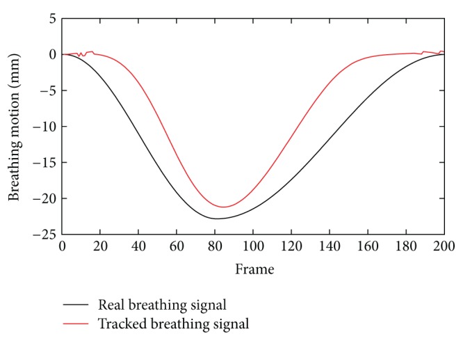
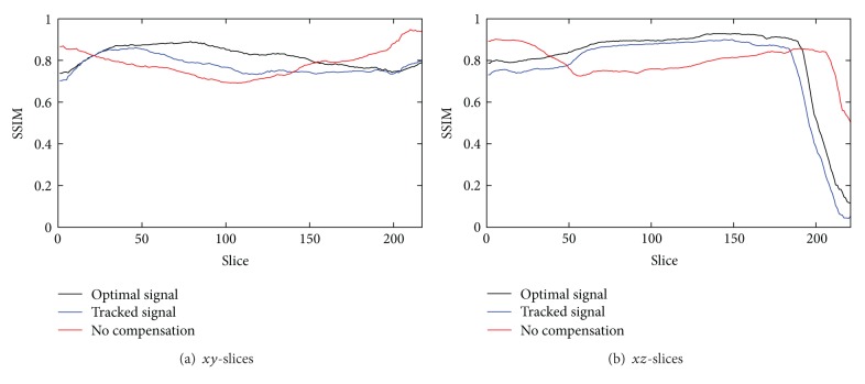
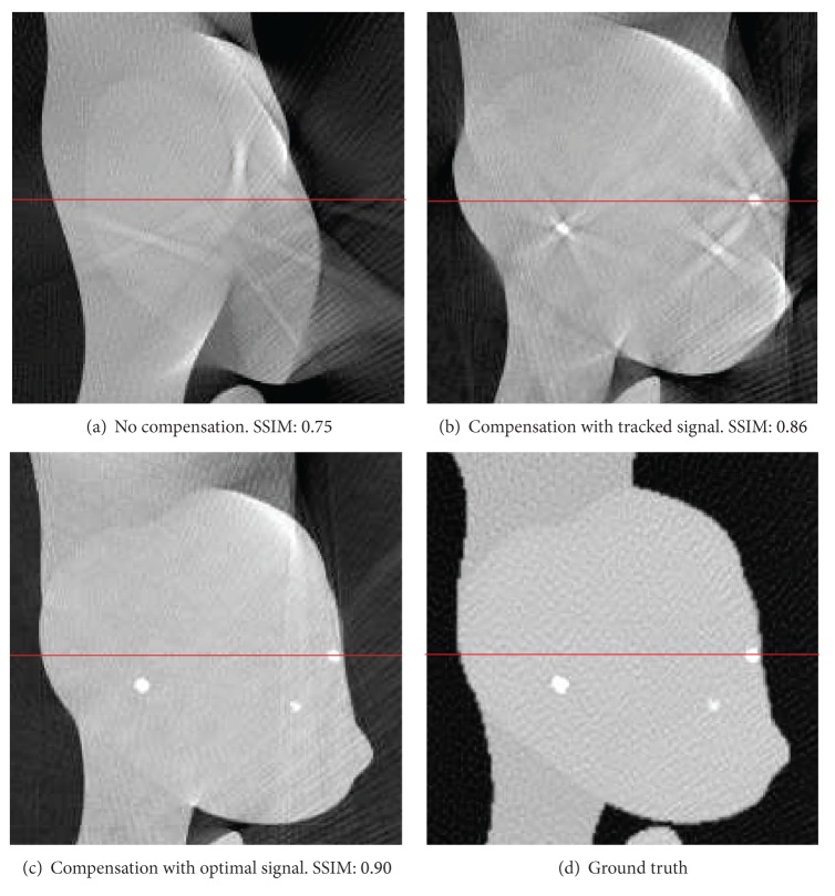
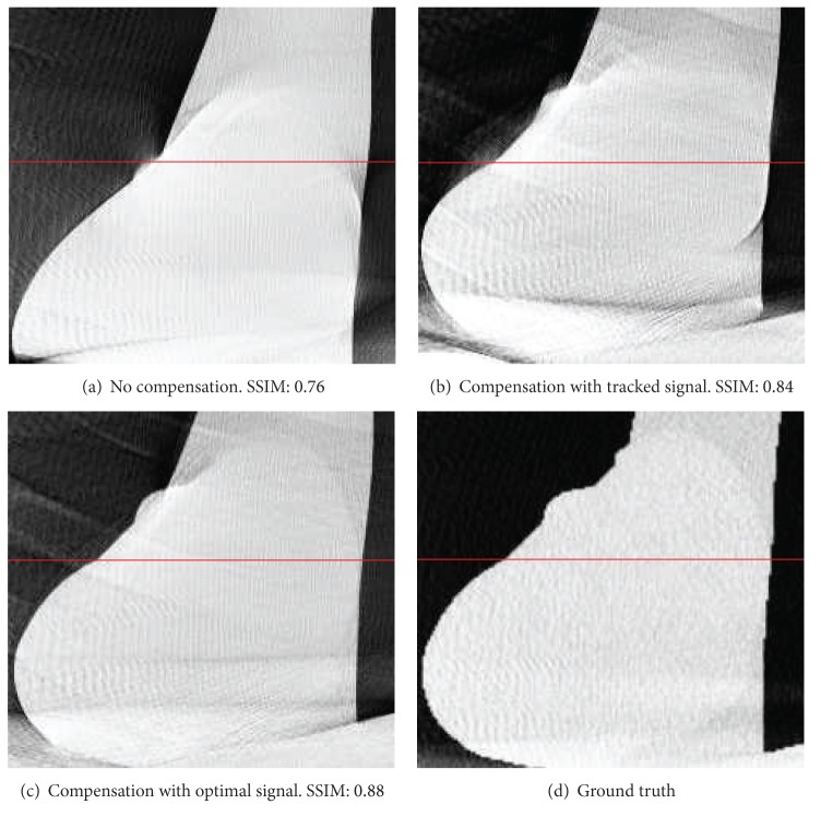
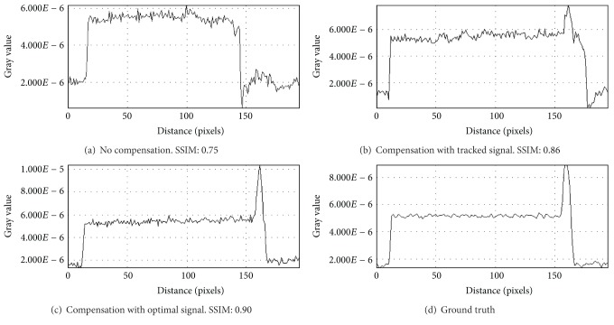
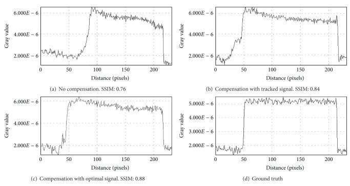
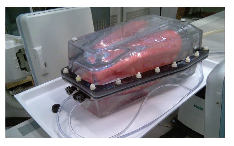
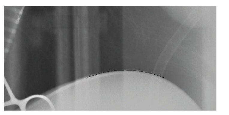
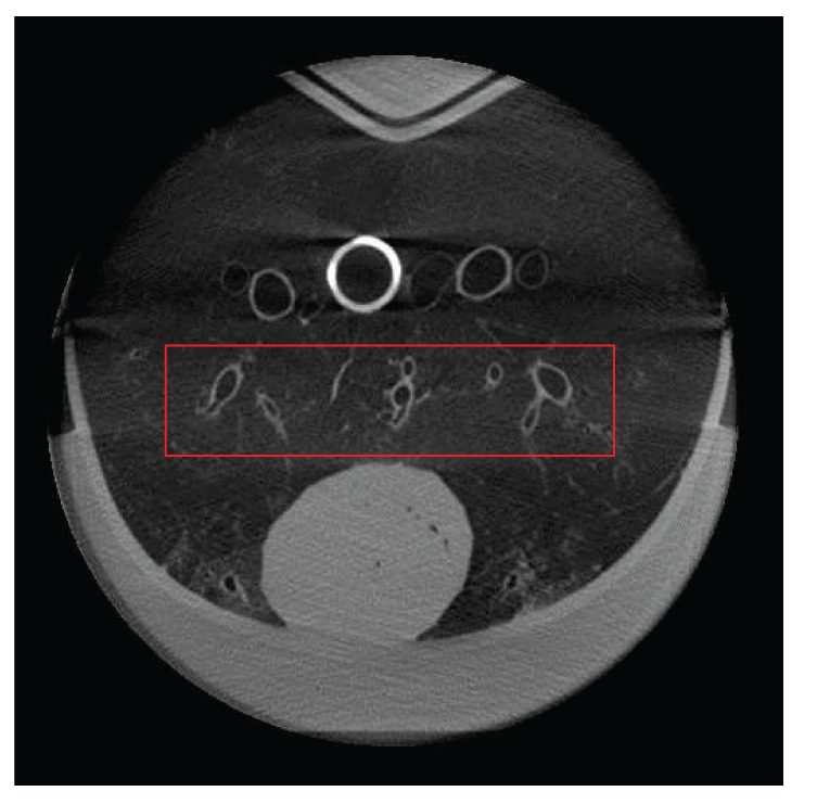
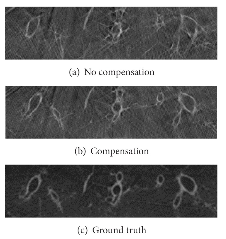
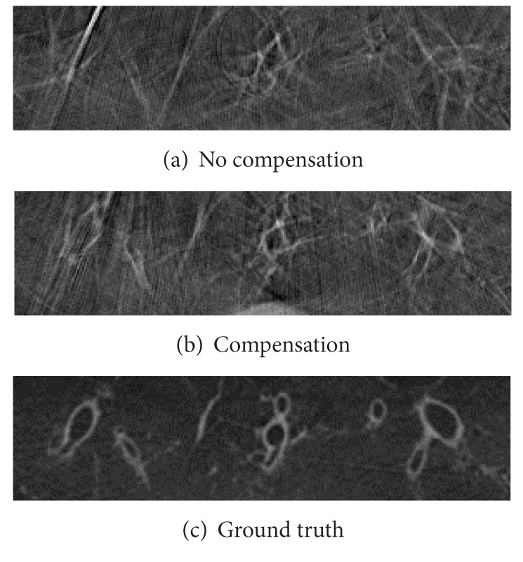
Long acquisition times lead to image artifacts in thoracic C-arm CT. Motion blur caused by respiratory motion leads to decreased image quality in many clinical applications. We introduce an image-based method to estimate and compensate respiratory motion in C-arm CT based on diaphragm motion. In order to estimate respiratory motion, we track the contour of the diaphragm in the projection image sequence. Using a motion corrected triangulation approach on the diaphragm vertex, we are able to estimate a motion signal. The estimated motion signal is used to compensate for respiratory motion in the target region, for example, heart or lungs. First, we evaluated our approach in a simulation study using XCAT. As ground truth data was available, a quantitative evaluation was performed. We observed an improvement of about 14% using the structural similarity index. In a real phantom study, using the artiCHEST phantom, we investigated the visibility of bronchial tubes in a porcine lung. Compared to an uncompensated scan, the visibility of bronchial structures is improved drastically. Preliminary results indicate that this kind of motion compensation can deliver a first step in reconstruction image quality improvement. Compared to ground truth data, image quality is still considerably reduced.
Figures
References
-
- Jahnke C, Paetsch I, Achenbach S, et al. Coronary MR imaging: breath-hold capability and patterns, coronary artery rest periods, and β-blocker use. Radiology. 2006;239(1):71–78. - PubMed
-
- Blondel C, Malandain G, Vaillant R, Ayache N. Reconstruction of coronary arteries from a single rotational X-ray projection sequence. IEEE Transactions on Medical Imaging. 2006;25(5):653–663. - PubMed
-
- Prümmer M, Hornegger J, Lauritsch G, Wigström L, Girard-Hughes E, Fahrig R. Cardiac c-arm CT: a unified framework for motion estimation and dynamic CT. IEEE Transactions on Medical Imaging. 2009;28(11):1836–1849. - PubMed
-
- Taguchi K, Segars WP, Fung GSK, Tsui BMW. Toward time resolved 4D cardiac CT imaging with patient dose reduction; estimating the global heart motion. Proceedings of the Medical Imaging 2006: Physics of Medical Imaging (SPIE '06); February 2006; San Diego, CA, USA. pp. 61 420J-1–61 420J-9.
-
- Schaller C, Penne J, Hornegger J. Time-of-flight sensor for respiratory motion gating. Medical Physics. 2008;35(7):3090–3093. - PubMed
LinkOut - more resources
Full Text Sources
Other Literature Sources
Research Materials
