

Systematic identification and characterization of novel human skin-associated genes encoding membrane and secreted proteins
- PMID: 23840300
- PMCID: PMC3688712
- DOI: 10.1371/journal.pone.0063949
Systematic identification and characterization of novel human skin-associated genes encoding membrane and secreted proteins
Abstract
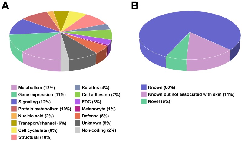
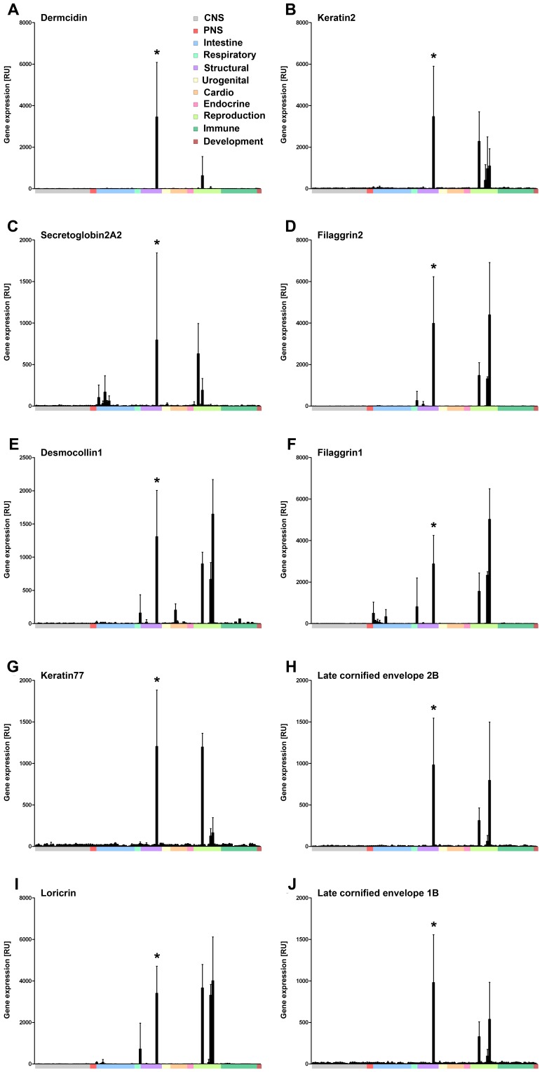
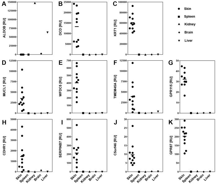
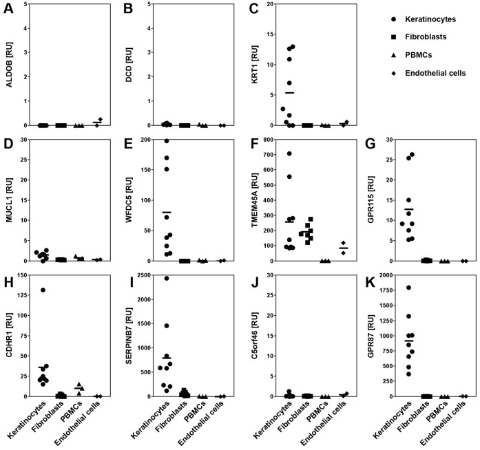
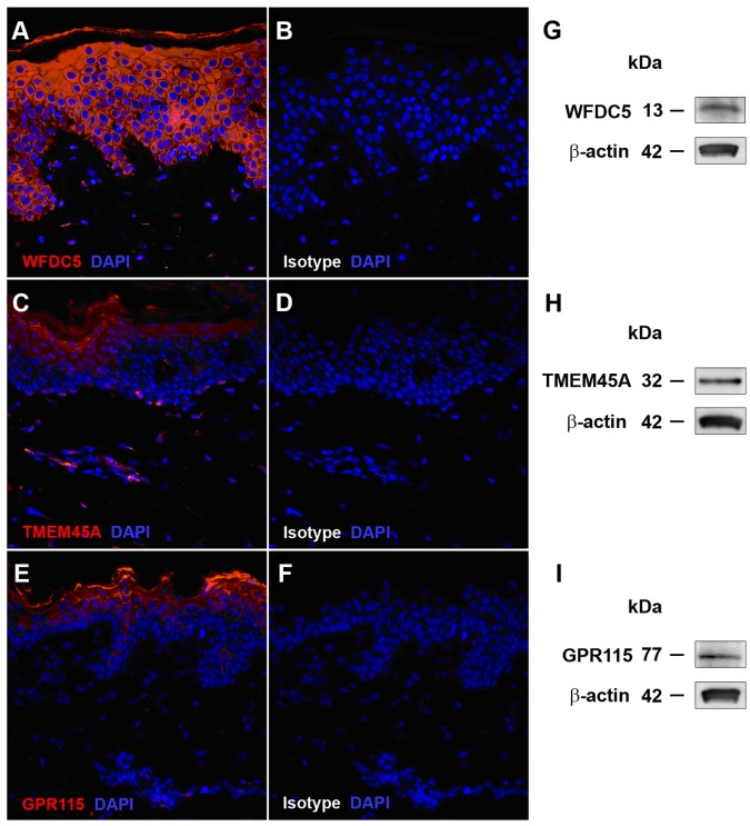
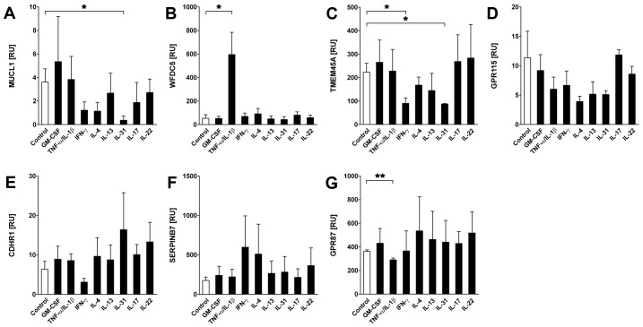
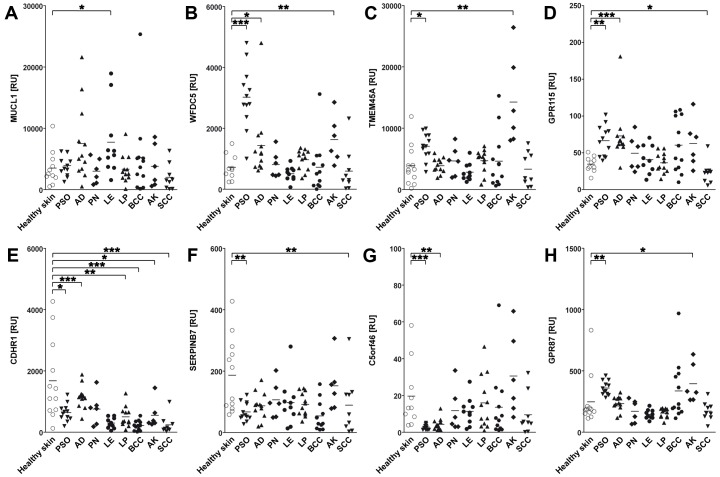
Through bioinformatics analyses of a human gene expression database representing 105 different tissues and cell types, we identified 687 skin-associated genes that are selectively and highly expressed in human skin. Over 50 of these represent uncharacterized genes not previously associated with skin and include a subset that encode novel secreted and plasma membrane proteins. The high levels of skin-associated expression for eight of these novel therapeutic target genes were confirmed by semi-quantitative real time PCR, western blot and immunohistochemical analyses of normal skin and skin-derived cell lines. Four of these are expressed specifically by epidermal keratinocytes; two that encode G-protein-coupled receptors (GPR87 and GPR115), and two that encode secreted proteins (WFDC5 and SERPINB7). Further analyses using cytokine-activated and terminally differentiated human primary keratinocytes or a panel of common inflammatory, autoimmune or malignant skin diseases revealed distinct patterns of regulation as well as disease associations that point to important roles in cutaneous homeostasis and disease. Some of these novel uncharacterized skin genes may represent potential biomarkers or drug targets for the development of future diagnostics or therapeutics.
Conflict of interest statement
Figures
Similar articles
-
Comprehensive RNA sequencing in primary murine keratinocytes and fibroblasts identifies novel biomarkers and provides potential therapeutic targets for skin-related diseases.Cell Mol Biol Lett. 2021 Oct 3;26(1):42. doi: 10.1186/s11658-021-00285-6. Cell Mol Biol Lett. 2021. PMID: 34602061 Free PMC article.
-
Systematic screening and identification of novel psoriasis‑specific genes from the transcriptome of psoriasis‑like keratinocytes.Mol Med Rep. 2019 Mar;19(3):1529-1542. doi: 10.3892/mmr.2018.9782. Epub 2018 Dec 20. Mol Med Rep. 2019. PMID: 30592269 Free PMC article.
-
RNA-Seq Analysis of IL-1B and IL-36 Responses in Epidermal Keratinocytes Identifies a Shared MyD88-Dependent Gene Signature.Front Immunol. 2018 Jan 29;9:80. doi: 10.3389/fimmu.2018.00080. eCollection 2018. Front Immunol. 2018. PMID: 29434599 Free PMC article.
-
LPA Induces Keratinocyte Differentiation and Promotes Skin Barrier Function through the LPAR1/LPAR5-RHO-ROCK-SRF Axis.J Invest Dermatol. 2019 May;139(5):1010-1022. doi: 10.1016/j.jid.2018.10.034. Epub 2018 Nov 14. J Invest Dermatol. 2019. PMID: 30447238 Review.
-
The top skin-associated genes: a comparative analysis of human and mouse skin transcriptomes.Biol Chem. 2014 Jun;395(6):577-91. doi: 10.1515/hsz-2013-0279. Biol Chem. 2014. PMID: 24497224 Review.
Cited by
-
Azithromycin induces epidermal differentiation and multivesicular bodies in airway epithelia.Respir Res. 2019 Jun 24;20(1):129. doi: 10.1186/s12931-019-1101-3. Respir Res. 2019. PMID: 31234850 Free PMC article.
-
TMEM45A Is Dispensable for Epidermal Morphogenesis, Keratinization and Barrier Formation.PLoS One. 2016 Jan 19;11(1):e0147069. doi: 10.1371/journal.pone.0147069. eCollection 2016. PLoS One. 2016. PMID: 26785122 Free PMC article.
-
Effect of Gentamicin Ointment in Patients with Nagashima-type Palmoplantar Keratosis: A Double-blind Vehicle-controlled Study.Acta Derm Venereol. 2021 Feb 11;101(2):adv00392. doi: 10.2340/00015555-3760. Acta Derm Venereol. 2021. PMID: 33554268 Free PMC article. Clinical Trial.
-
GPR115 Contributes to Lung Adenocarcinoma Metastasis Associated With LAMC2 and Predicts a Poor Prognosis.Front Oncol. 2020 Nov 20;10:577530. doi: 10.3389/fonc.2020.577530. eCollection 2020. Front Oncol. 2020. PMID: 33330053 Free PMC article.
-
TMEM45A, SERPINB5 and p16INK4A transcript levels are predictive for development of high-grade cervical lesions.Am J Cancer Res. 2016 Jul 1;6(7):1524-36. eCollection 2016. Am J Cancer Res. 2016. PMID: 27508094 Free PMC article.
References
-
- Blumenberg M (2006) DNA microarrays in dermatology and skin biology. OMICS 10: 243–260. - PubMed
-
- Curto EV, Lambert GW, Davis RL, Wilborn TW, Dooley TP (2002) Biomarkers of human skin cells identified using DermArray DNA arrays and new bioinformatics methods. Biochem Biophys Res Commun 291: 1052–1064. - PubMed
-
- Haider AS, Lowes MA, Suarez-Farinas M, Zaba LC, Cardinale I, et al. (2008) Cellular genomic maps help dissect pathology in human skin disease. J Invest Dermatol 128: 606–615. - PubMed
-
- Rinn JL, Wang JK, Liu H, Montgomery K, van de Rijn M, et al. (2008) A systems biology approach to anatomic diversity of skin. J Invest Dermatol 128: 776–782. - PubMed
Publication types
MeSH terms
Substances
Grants and funding
LinkOut - more resources
Full Text Sources
Other Literature Sources
