. 2013 Jun 19;8(6):e66207.
doi: 10.1371/journal.pone.0066207. Print 2013.

A Low Temperature Limit for Life on Earth

Affiliations

A Low Temperature Limit for Life on Earth

Andrew Clarke et al. PLoS One. .

Abstract

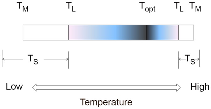
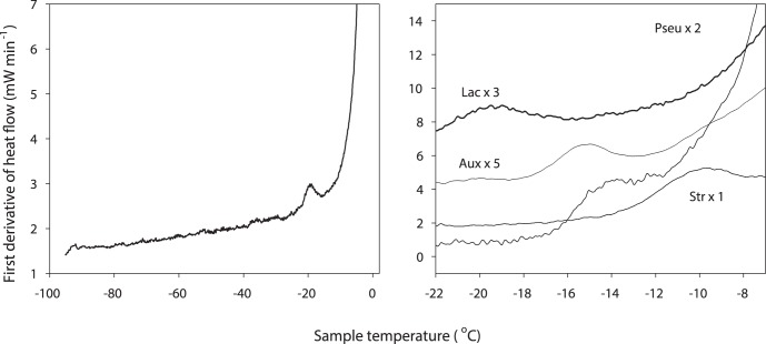
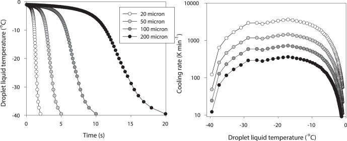
There is no generally accepted value for the lower temperature limit for life on Earth. We present empirical evidence that free-living microbial cells cooling in the presence of external ice will undergo freeze-induced desiccation and a glass transition (vitrification) at a temperature between -10°C and -26°C. In contrast to intracellular freezing, vitrification does not result in death and cells may survive very low temperatures once vitrified. The high internal viscosity following vitrification means that diffusion of oxygen and metabolites is slowed to such an extent that cellular metabolism ceases. The temperature range for intracellular vitrification makes this a process of fundamental ecological significance for free-living microbes. It is only where extracellular ice is not present that cells can continue to metabolise below these temperatures, and water droplets in clouds provide an important example of such a habitat. In multicellular organisms the cells are isolated from ice in the environment, and the major factor dictating how they respond to low temperature is the physical state of the extracellular fluid. Where this fluid freezes, then the cells will dehydrate and vitrify in a manner analogous to free-living microbes. Where the extracellular fluid undercools then cells can continue to metabolise, albeit slowly, to temperatures below the vitrification temperature of free-living microbes. Evidence suggests that these cells do also eventually vitrify, but at lower temperatures that may be below -50°C. Since cells must return to a fluid state to resume metabolism and complete their life cycle, and ice is almost universally present in environments at sub-zero temperatures, we propose that the vitrification temperature represents a general lower thermal limit to life on Earth, though its precise value differs between unicellular (typically above -20°C) and multicellular organisms (typically below -20°C). Few multicellular organisms can, however, complete their life cycle at temperatures below ∼-2°C.


Conflict of interest statement

Competing Interests: The authors have the following interests. G. John Morris receives a salary from Asymptote. The work described in the paper was entirely without the activities of Asymptote. This does not alter the authors’ adherence to all the PLOS ONE policies on sharing data and materials.

Figures

Figure 1
Figure 1. Temperature limits for life.
TL: temperature limit for completion of the life cycle, TM: temperature limit for metabolism, TS: temperature limit for survival. Note that TS can be above, at, or below TM. The shaded area shows temperature range over which the organism can complete its life cycle, and the white areas show the temperature range (upper and lower) for survival. Topt is the temperature at which growth rate is maximal, which is typically closer to the upper TL than the lower TL.
Figure 2
Figure 2. Structure of ice and cells following cooling of a cell suspension.
Cryo scanning electron microscopy of fracture samples of Lactobacillus delbrueckii ssp. bulgaricus following slow cooling. A. Low power image of a fractured sample, showing cells confined to the unfrozen liquid between ice crystals; this unfrozen matrix is highly freeze-concentrated as solutes are excluded from the growing ice crystal. B. High power image of the interface between ice and the freeze concentrated matrix, with bacterial cells labelled. C. Freeze substitution of the same sample reveals cells packed within the freeze concentrated matrix. In all samples the spaces originally occupied by ice crystals are revealed as voids following sublimation of ice. The labels mark ice voids (*) Lactobacillus cells (Lb) and freeze concentrated solute (+).
Figure 3
Figure 3. Differential scanning calorimetry (DSC) traces showing vitrification in cell suspensions.
A (left panel). DSC trace of Lactobacillus delbrueckii ssp. bulgaricus which clearly shows a vitrification (colloid glass transition) temperature, Tg, of −19.3°C in samples that have been cooled below −90°C and then warmed. The glass transition during warming is indicated by the deviation in the first derivative of the heat flow, associated with a change in heat capacity as the bacterial cells devitrify at Tg. The large increase in heat flow at temperatures above Tg corresponds to the freezing of the aqueous medium in which the cells were suspended. B (right panel). Composite of DSC traces from four organisms (three bacteria, and an alga). The traces have been scaled as shown to render them visible on a single ordinate scale.
Figure 4
Figure 4. Temperature profile through an ice core taken at Dome C, Antarctica.
Data are averaged over bins of 5 m. The dotted lines show a putative threshold depth above which cells will be vitrified and hence not metabolising, and below which cells may be in a fluid state and able to metabolise, albeit slowly. At the bottom of the ice core the temperature may be sufficient for cell growth and division to be possible, assuming the presence of sufficient free energy (suitable electron donors and acceptors) and a source of nutrients. Core temperature data courtesy of the European Project for Ice Coring in Antarctica, EPICA.
Figure 5
Figure 5. Temperature following nucleation of water droplets of different sizes.
A (left panel). Temperature of cloud droplets following ice nucleation at an environmental temperature of −40°C modelled for droplets of diameter 20 µm (left hand trace), 50 µm, 100 µm and 200 µm (right hand trace). The traces show the cooling phase after release of latent heat has raised droplet temperature from −40°C to almost 0°C. For all droplets cooling rates exceed 100 K min−1 and for the smaller sizes exceed 1000 K min−1. B (right panel). Cooling rate (K min−1, log scale) for these droplets, showing the very fast cooling rates following nucleation at −40°C in small droplets; these rates are sufficient to induce lethal intracellular freezing in any cells contained within the droplet.

References

    1. Kashefi K, Lovley DR (2003) Extending the upper temperature limit for life. Science 301(5635): 934–934. - PubMed
    1. Pace NR (2001) The universal nature of biochemistry. Proceedings of the National Academy of Sciences of the United States of America 98(3): 805–808. - PMC - PubMed
    1. Kerry KR, Grace DR, Williams R, Burton HR (1977) Studies on some saline lakes of the Vestfold Hills, Antarctica. In: Llano GA, editor. Adaptations within Antarctic ecosystems. Washington, D.C.: Smithsonian Institution. 839–858.
    1. Tominaga H, Fukui F (1981) Saline lakes in Syowa Oasis, Antarctica. Hydrobiologia 82(3): 375–389.
    1. Murray AE, Kenig F, Fritsen CH, McKay CP, Cawley KM, et al. (2012) Microbial life at −13°C in the brine of an ice-sealed Antarctic lake. Proceedings of the National Academy of Sciences of the United States of America 109(50): 20626–20631. - PMC - PubMed