. 2013 Jun 19;8(6):e67076.
doi: 10.1371/journal.pone.0067076. Print 2013.

The ArfGEF GBF-1 Is Required for ER Structure, Secretion and Endocytic Transport in C. elegans

Affiliations

The ArfGEF GBF-1 Is Required for ER Structure, Secretion and Endocytic Transport in C. elegans

Karin B Ackema et al. PLoS One. .

Abstract

Small GTPases of the Sar/Arf family are essential to generate transport containers that mediate communication between organelles of the secretory pathway. Guanine nucleotide exchange factor (GEFs) activate the small GTPases and help their anchorage in the membrane. Thus, GEFs in a way temporally and spatially control Sar1/Arf1 GTPase activation. We investigated the role of the ArfGEF GBF-1 in C. elegans oocytes and intestinal epithelial cells. GBF-1 localizes to the cis-Golgi and is part of the t-ER-Golgi elements. GBF-1 is required for secretion and Golgi integrity. In addition, gbf-1(RNAi) causes the ER reticular structure to become dispersed, without destroying ER exit sites (ERES) because the ERES protein SEC-16 was still localized in distinct punctae at t-ER-Golgi units. Moreover, GBF-1 plays a role in receptor-mediated endocytosis in oocytes, without affecting recycling pathways. We find that both the yolk receptor RME-2 and the recycling endosome-associated RAB-11 localize similarly in control and gbf-1(RNAi) oocytes. While RAB5-positive early endosomes appear to be less prominent and the RAB-5 levels are reduced by gbf-1(RNAi) in the intestine, RAB-7-positive late endosomes were more abundant and formed aggregates and tubular structures. Our data suggest a role for GBF-1 in ER structure and endosomal traffic.


Conflict of interest statement

Competing Interests: The authors have declared that no competing interests exist.

Figures

Figure 1
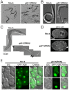
Figure 1. gbf-1(RNAi) worms causes multiple development defects.
(A) gbf-1(RNAi) worms have a weakened cuticle. When fed from hatching on gbf-1(RNAi) bacteria, the majority of the worms died of cuticle bursting upon reaching adulthood. Adult worms and their offspring on agar plates. Black arrows point to worms that have bursted. This phenotype is consistently seen in all gbf-1(RNAi) feedings from hatching (B) Although most embryos arrested early in development in gfb-1(RNAi) worms, few larvae could hatch from the eggs. These larvae often displayed severe problems with their cuticle formation. Black arrow points to an intestine that is directly exposed to the environment instead of being protected by the cuticle. (C) Worms fed on gbf-1(RNAi) from hatching frequently accumulate liquid filled vacuoles in the body cavity. Furthermore they are sterile (no eggs in the uterus) and their intestine appears ‘clear’. Merged bright field images of whole worms. Black arrows point to vacuoles. (D) During meiosis the egg normally expels two polar bodies during anaphase I and II. In gbf-1(RNAi) eggs, the extrusion of the second polar body during anaphase II often failed, which can be seen by the presence of 3 nuclei (arrow) in fertilized eggs at pronuclear meeting. This phenotype was consistently seen throughout different experiments involving embryos. We used SP12::GFP marking the ER that nicely outlined the nuclei. Anterior in both one-cell embryos is on the left. A single plane confocal image is shown. (E) Offspring from worms fed on gbf-1(RNAi) from L3 frequently show problems with cytokinesis which results in unequal distribution of the DNA between cells. GFP::H2B was used to visualize the DNA. The experiment was performed at least three times.
Figure 2
Figure 2. GBF-1 is important for Golgi integrity.
(A) GBF-1 localizes to the Golgi in oocytes. Displayed are the three most proximal oocytes. GBF-1 partially co-localized with the medial Golgi marker UGTP-1::GFP. The third panel shows an overlay of the two channels and an enlargement of the white box is shown on the right. (B) GBF-1 is part of the Golgi t-ER region in oocytes. GBF-1 was localized juxtaposed to the ERES marker SEC-16. (C) The Golgi in C. elegans germ line is organized in small mini stacks localized in close proximity to ER-exit sites (ERES). GBF-1 was found ‘sandwiched’ between the Golgi marker UGTP::GFP and ERES marker SEC-16. (D) agef-1(RNAi) and gbf-1(RNAi) causes the collapse of the Golgi around the nuclear membrane and cell periphery in oocytes. A single confocal plane in the center of the cell is shown. (A–D) The orientation of the gonad is identical in all panels: the spermatheca and the most mature oocyte are located to the left. All channels are in false colors. A single confocal plane is shown. (E) Transmission electron microscopy images of oocytes. The top panel shows a part of the oocytes. The bottom panel shows an enlargement of a Golgi stack as indicated by a red box. Largely inflated cisternae were observed in both agef-1(RNAi) and gbf-1(RNAi) cells as indicated by asterisks. gbf-1(RNAi) Golgi stacks accumulated a lot of small-sized vesicles as indicated by black arrows. (A–E) The experiments have been performed at least three times with comparable experimental conditions using different RNAi feeding batches. The selected images are representative for the observed phenotype.
Figure 3
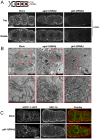
Figure 3. GBF-1 is required for ER integrity in oocytes.
(A) gbf-1(RNAi) results in a disorganized ER in oocytes. The ER appeared dispersed in gbf-1(RNAi). ER in the gonad was stained with an anti-HDEL antibody. Two focal planes are shown, one at the level of the nuclei (’Middle’) and one at the cell cortex (‘Top’) as indicated by the diagram. The spermatheca is located to the left. In wild type oocytes, the ER was organized in a weakly reticulated structure distributed throughout the cytoplasm. The ER morphology in agef-1(RNAi) oocytes was not affected, while in gbf-1(RNAi) oocytes the ER was present as dispersed haze. (B) TEM images of oocytes. The top panel shows a part of an oocyte and the bottom panel an enlargement as indicated by the red box. The ER in wild-type and agef-1(RNAi) oocytes was present in regular reticulate structures, indicated by ‘ER’. In gbf-1(RNAi) the ER was disorganized with regions where the lumen was collapsed. V, vesicle; M, mitochondria; G, Golgi; N, nucleus. (C) ERES are formed normally in gbf-1(RNAi) oocytes. Immunofluorescence images of oocytes which were stained with UGTP::GFP and anti-SEC-16. In a blind experiment, the SEC-16 staining in gbf-1(RNAi) and mock treated oocytes was indistinguishable from each other. Pictures refer to a single confocal plane. The spermatheca is located to the left. (A–C) The experiments have been performed at least three times using different RNAi feeding batches under the same experimental conditions. The selected images are representative for the observed phenotype.
Figure 4
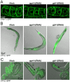
Figure 4. GBF-1 is required for secretion and endocytosis.
(A) GBF-1 and AGEF-1 are required for caveolin secretion in oocytes. In both agef-1(RNAi) and gbf-1(RNAi) oocytes less CAV-1::GFP vesicles were observed throughout the cell when compared to mock treated animals. Single confocal planes are shown. (B) Endocytosis of yolk protein is impaired in gbf-1(RNAi) worms. In mock-treated and agef-1(RNAi) worms, YP170::GFP (VIT-2::GFP) accumulated in the maturing oocytes and in the developing embryos. In gbf-1(RNAi) worms a strong accumulation of YP170::GFP in the body cavity was observed in approximately 70% of the worms. (C) AGEF-1 and GBF-1 are required for efficient secretion of yolk protein. Both agef-1(RNAi) and gbf-1(RNAi) caused a buildup of yolk (YP170::GFP) in the intestine (indicated by black arrows). A DIC with GFP overlay is shown. (A–C) All experiments have been performed at least three times using similar experimental conditions using different RNAi feeding batches. The selected images are representative for the observed phenotype.
Figure 5
Figure 5. Recycling and internalization is not affected in gbf-1(RNAi) worms.
(A) Yolk protein uptake is strongly reduced in gbf-1(RNAi) oocytes. A Single confocal plane through the center of the cells is shown. The spermatheca and the most mature oocyte are oriented toward the top. (B) Localization and intensity of the yolk receptor RME-2::GFP in gbf-1(RNAi) oocytes is undistinguishable from that in control worms. An intensity plot is shown below their corresponding images. The peaks represent the fluorescence at the plasma membrane between cell boundaries. Gray values are given in arbitrary units. (C) Recycling to the plasma membrane is unaffected in gbf-1(RNAi) oocytes. RAB11.1::GFP labeled recycling endosomes reached the plasma membrane normally in gbf-1(RNAi) oocytes compared to control oocytes. A single confocal plane is shown. (D) Knockdown of GBF-1 does not alter endocytosis or degradation of caveolin. Internalization of CAV-1::GFP in gbf-1(RNAi) oocytes was indistinguishable from that in mock RNAi worms. Spermatheca is located to the left. (E) RME-2 is endocytosed and degraded in gbf-1(RNAi) one-cell embryos. The time required for internalization of RME-2 in gbf-1(RNAi) oocytes seems not to be different from that observed in mock RNAi control worms. Anterior of the embryo is oriented towards the top. Corresponding fluorescence and DIC images are shown. (A–E) All experiments have been performed at least three times with comparable experimental conditions using different RNAi feeding batches. The selected images are representative for the observed phenotype.
Figure 6
Figure 6. GBF-1 plays a role in later stages of endocytosis.
(A) The endocytic pathway in the gbf-1(RNAi) intestine is severely disturbed. A schematic drawing of the intestine is shown. The expression of the early endocytic marker RAB-5::GFP was strongly reduced, while late endosomes appeared to form large aggregates as indicated by the presence of RAB-7. These phenotypes were observed in approximately 60%–90% of gbf-1(RNAi) worms compared to ≥10% in mock treated worms. The experiment was performed four times with 15 worms in each experiment. Single plane confocal images are displayed. (B) GBF-1 and AGEF-1 are both required for correct secretion from the Golgi. GBF-1 mainly acts at the cis-Golgi, while AGEF-1 is more important at the trans-Golgi. GBF-1 is involved in the endocytic pathway in C. elegans. Our data suggest a function at early and late endosomes, while in Drosophila GBF-1 was required at the plasma membrane.

References

    1. Poteryaev D, Datta S, Ackema K, Zerial M, Spang A (2010) Identification of the switch in early-to-late endosome transition. Cell 141: 497–508. - PubMed
    1. Morinaga N, Tsai SC, Moss J, Vaughan M (1996) Isolation of a brefeldin A-inhibited guanine nucleotide-exchange protein for ADP ribosylation factor (ARF) 1 and ARF3 that contains a Sec7-like domain. Proceedings of the National Academy of Sciences of the United States of America 93: 12856–12860. - PMC - PubMed
    1. Sato K, Sato M, Audhya A, Oegema K, Schweinsberg P, et al. (2006) Dynamic regulation of caveolin-1 trafficking in the germ line and embryo of Caenorhabditis elegans. Mol Biol Cell 17: 3085–3094. - PMC - PubMed
    1. Peyroche A, Paris S, Jackson CL (1996) Nucleotide exchange on ARF mediated by yeast Gea1 protein. Nature 384: 479–481. - PubMed
    1. Spang A, Herrmann JM, Hamamoto S, Schekman R (2001) The ADP ribosylation factor-nucleotide exchange factors Gea1p and Gea2p have overlapping, but not redundant functions in retrograde transport from the Golgi to the endoplasmic reticulum. Mol Biol Cell 12: 1035–1045. - PMC - PubMed

MeSH terms

Substances

LinkOut - more resources