. 2013 Jun 26;8(6):e67266.
doi: 10.1371/journal.pone.0067266. Print 2013.

Igf Signaling is Required for Cardiomyocyte Proliferation during Zebrafish Heart Development and Regeneration

Affiliations

Igf Signaling is Required for Cardiomyocyte Proliferation during Zebrafish Heart Development and Regeneration

Ying Huang et al. PLoS One. .

Abstract

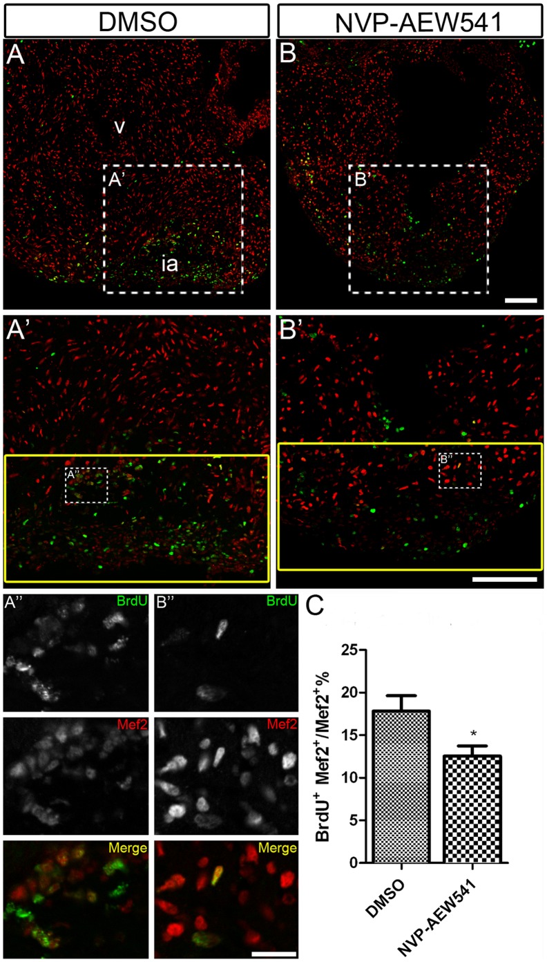
Unlike its mammalian counterpart, the adult zebrafish heart is able to fully regenerate after severe injury. One of the most important events during the regeneration process is cardiomyocyte proliferation, which results in the replacement of lost myocardium. Growth factors that induce cardiomyocyte proliferation during zebrafish heart regeneration remain to be identified. Signaling pathways important for heart development might be reutilized during heart regeneration. IGF2 was recently shown to be important for cardiomyocyte proliferation and heart growth during mid-gestation heart development in mice, although its role in heart regeneration is unknown. We found that expression of igf2b was upregulated during zebrafish heart regeneration. Following resection of the ventricle apex, igf2b expression was detected in the wound, endocardium and epicardium at a time that coincides with cardiomyocyte proliferation. Transgenic zebrafish embryos expressing a dominant negative form of Igf1 receptor (dn-Igf1r) had fewer cardiomyocytes and impaired heart development, as did embryos treated with an Igf1r inhibitor. Moreover, inhibition of Igf1r signaling blocked cardiomyocyte proliferation during heart development and regeneration. We found that Igf signaling is required for a subpopulation of cardiomyocytes marked by gata4:EGFP to contribute to the regenerating area. Our findings suggest that Igf signaling is important for heart development and myocardial regeneration in zebrafish.


Conflict of interest statement

Competing Interests: The authors have declared that no competing interests exist.

Figures

Figure 1
Figure 1. igf2b is upregulated during zebrafish heart regeneration.
In situ hybridization (ISH) was performed on sham operated hearts (A) and 3 dpa (B), 7 dpa (C), 10 dpa (D) and 14 dpa (E) regenerating hearts. There is no detectable igf2b expression in sham-operated hearts by ISH, whereas strong expression is induced in endocardium (arrows in B and inset B’) and epicardium (arrows in C and inset C’) near the wound following amputation of the ventricular apex. Red dashed lines mark the amputation site and wound area. v: ventricle, ia: injured area, epi: epicardium. Scale bar = 100 µm. (F) Quantitative RT-PCR of igf 2b expression at 3, 7, 10 and 14 dpa compared to uncut hearts.
Figure 2
Figure 2. Igf signaling is required for proper cardiomyocyte number in zebrafish embryonic hearts.
The number of ventricular cardiomyocytes (area encircled by dotted line) number decreased in embryos when Igf1r signaling was blocked. (A) Tg(myl7:nDsRed) single transgenic and (C) Tg(hsp:dnigf1ra-GFP; myl7:nDsRed) double transgenic embryos (n = 16) were heat shocked at 40°C for 30 min around 48 and 72 hpf and observed at 76 hpf. dnigf1ra embryos can be identified by GFP expression (C’ inset). Tg(myl7:nDsRed) single transgenic siblings (n = 16) were used as controls. myl7:nDsRed embryos were treated with Igf1r inhibitor (D) (n = 20) and DMSO as a control (B) (n = 20) from 48 to 72 hpf and observed at 76 hpf. a: atrium, v: ventricle. Scale bar = 20 µm. (E, F) Quantification of mean ventricular cardiomyocyte number ± S.E. (***p<0.0001; **p<0.001). Fewer ventricular cardiomyocytes were observed around 76 hpf after Igf1r signaling was blocked.
Figure 3
Figure 3. Igf signaling is required for cardiomyocyte proliferation during zebrafish heart development.
Wild type fish were treated with DMSO as a control (n = 5) (A and A’) and the Igf1r inhibitor NVP-AEW541 (n = 7) (B and B’) from 48–72 hpf. BrdU was added for the same time period. BrdU (green) and Mef2 (red) double positive cells indicate proliferating cardiomyocytes (A, A’, B, B’). A’ and B’ are images of the dashed boxes in A and B. (A’ and B’), BrdU (green) and Mef2 (red) staining were shown as black and white or merged color images. Scale bar: (B) = 50 µm, (B’) = 20 µm. v: ventricle. (C) Quantification of BrdU positive cardiomyocytes (Mef2 positive) ± S. E. A significant decrease (***p<0.0001) in cardiomyocyte proliferation was detected in embryos treated with NVP-AEW541.
Figure 4
Figure 4. Igf signaling is required for cardiomyocyte proliferation during zebrafish heart regeneration.
Cardiomyocyte proliferation was decreased after Igf signaling was blocked during zebrafish heart regeneration. Tg(hsp70:gal4) control (n = 5) (A, A’, A’’) and Tg(hsp70:dnigf1ra-GFP) transgenic zebrafish (n = 7) (B, B’,B’’) zebrafish were heat shocked for 1 h at 38°C after amputation from 2–10 dpa. BrdU (green) and Mef2 (red) double positive cells indicate proliferating cardiomyocytes (A, A’, B, B’). A’ and B’ are the higher magnification images of the dashed boxes in A and B. A’’ and B’’ are the higher magnification images of the dashed boxes in A’ and B’. The yellow box indicates the wound area; cardiomyocytes were counted in this region. (A’’ and B’’), BrdU (green) and Mef2 (red) staining were shown as separated channel images (black and white) or merged color images. ia: injured area, v: ventricle. Scale Bar: (B, B’) = 100 µm, (B’’) = 20 µm. (C) Quantification of BrdU positive cardiomyocytes (Mef2 positive) ± S.E. A significant decrease (*p<0.05) in cardiomyocyte proliferation was detected in Tg(hsp:dnigf1ra-GFP) fish.
Figure 5
Figure 5. Chemical inhibition of Igf signaling suppresses cardiomyocyte proliferation during zebrafish heart regeneration.
Wild type fish were treated with DMSO as a control (A, A’ and A’’) (n = 5) and the Igf1r inhibitor NVP-AEW541 (B, B’ and B’’) (n = 5) from 2–14 dpa. BrdU (green) and Mef2 (red) double positive cells indicate proliferating cardiomyocytes (A, A’, B, B’). A’ and B’ are the higher magnification images of the dashed boxes in A and B. A’’ and B’’ are the higher magnification images of the dashed boxes in A’ and B’. The yellow box indicates the wound area and cardiomyocytes were counted in this region. (A’’ and B’’), BrdU (green) and Mef2 (red) staining were shown as black and white or merged color images. Scale bar: (B, B’) = 100 µm, (B’’) = 20 µm. ia: injured area, v: ventricle. (C) Quantification of BrdU positive cardiomyocytes (Mef2 positive) ± S.E. A significant decrease (*p<0.01) in cardiomyocyte proliferation was detected in fish treated with Igf1r inhibitor NVP-AEW541.
Figure 6
Figure 6. Igf signaling is required for heart regeneration.
Wild type fish were treated DMSO (A) (n = 6) or the Igf1r inhibitor NVP-AEW541 (n = 7) (B and C) from 2–30 dpa. AFOG staining was performed at 30 dpa to detect collagen (blue) and fibrin (red) deposition. The dashed line marks the regenerating area. Scale bar = 100 µm. ia: injured area. (D) Quantification of scar area normalized to ventricle area in DMSO and NVP-AEW541 treated hearts. A significant increase (*p<0.05) in scar/ventricular area ratio was detected in NVP-AEW541 treated hearts.
Figure 7
Figure 7. Igf signaling is required for contribution ofgata4:EGFP positive cardiomyocytes to the regenerating area.
gata4:EGFP fish were treated with the Igf1r inhibitor NVP-AEW541 from 2–14 dpa (n = 12) (B, D) and 7–10 dpa (n = 5) (F, H). DMSO was used as a control (14dpa: n = 10; 10 dpa: n = 6) (A, C, E, G). Whole mount confocal microscopy from the view of the apex (A, B, E, F) and frozen sections (C, D, G, H) were performed at 14 and 10 dpa to determine the contribution of the EGFP positive population. BrdU (red) and Gata4 (green) double positive cells indicate proliferating gata4:EGFP positive cardiomyocytes (G, G’, H, H’). G’, and H’ are the higher magnification images of the dashed boxes in G and H. BrdU staining (red) and gata4:EGFP (green) were shown as separated channel images. The dashed line marks the regenerating area. Scale bar: (B, D, F, H) = 50 µm; (H’) = 20 µm. ia: injured area, v: ventricle. (I) Quantification of BrdU and gata4:EGFP double positive cells/gata4:EGFP area ± S.E. A significant decrease (*p<0.05) in gata4:EGFP positive cell proliferation was detected in fish treated with Igf1r inhibitor NVP-AEW541 from 7–10dpa.

References

    1. Li F, Wang X, Capasso JM, Gerdes AM (1996) Rapid transition of cardiac myocytes from hyperplasia to hypertrophy during postnatal development. J Mol Cell Cardiol 28: 1737–1746. - PubMed
    1. Pasumarthi KB, Field LJ (2002) Cardiomyocyte cell cycle regulation. Circ Res 90: 1044–1054. - PubMed
    1. van Amerongen MJ, Engel FB (2008) Features of cardiomyocyte proliferation and its potential for cardiac regeneration. J Cell Mol Med 12: 2233–2244. - PMC - PubMed
    1. Walsh S, Ponten A, Fleischmann BK, Jovinge S (2010) Cardiomyocyte cell cycle control and growth estimation in vivo–an analysis based on cardiomyocyte nuclei. Cardiovasc Res 86: 365–373. - PubMed
    1. Roger VL, Go AS, Lloyd-Jones DM, Benjamin EJ, Berry JD, et al. (2012) Heart disease and stroke statistics–2012 update: a report from the American Heart Association. Circulation 125: e2–e220. - PMC - PubMed

Publication types