. 2013 Jun 28;8(6):e67303.
doi: 10.1371/journal.pone.0067303. Print 2013.

HGF mediates the anti-inflammatory effects of PRP on injured tendons

Affiliations

HGF mediates the anti-inflammatory effects of PRP on injured tendons

Jianying Zhang et al. PLoS One. .

Abstract

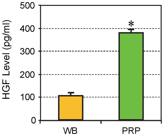
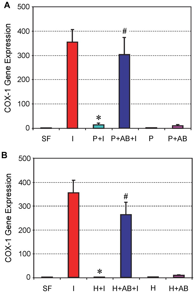
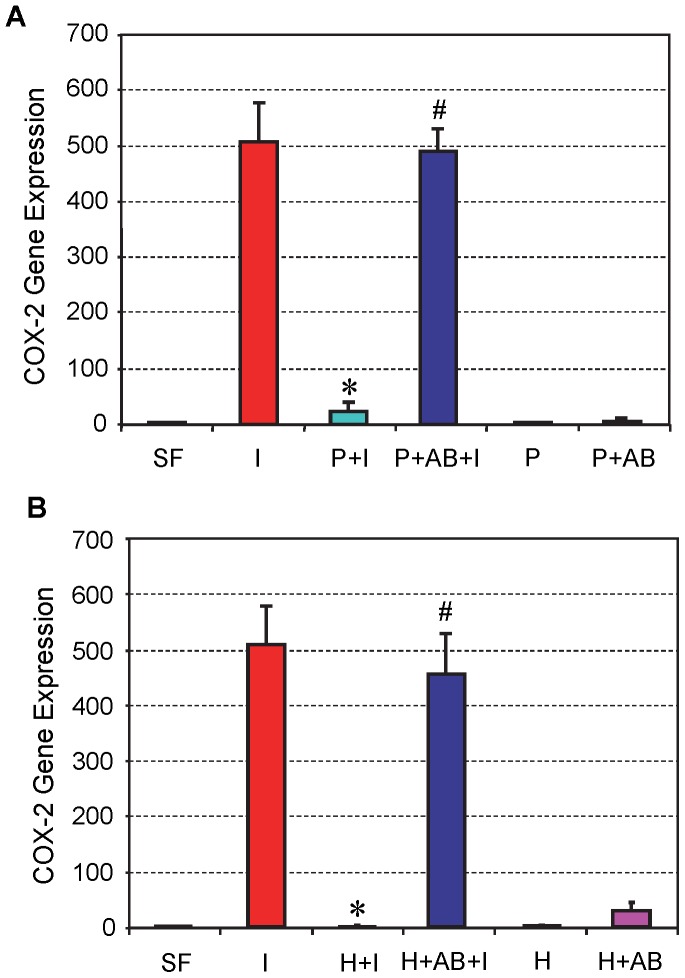
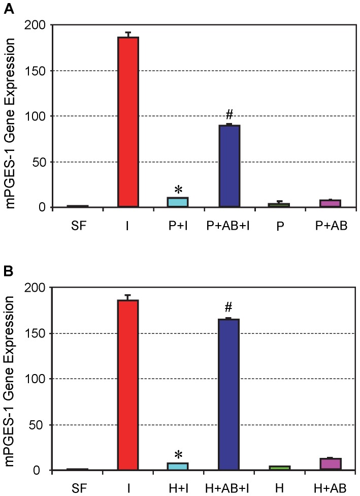
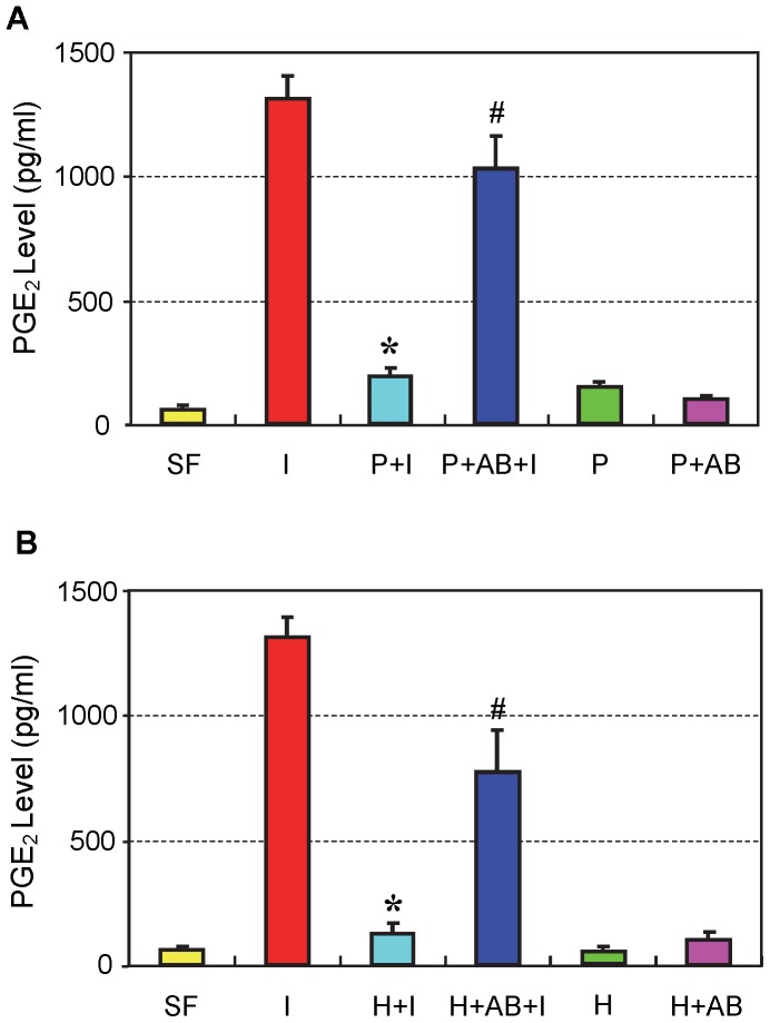
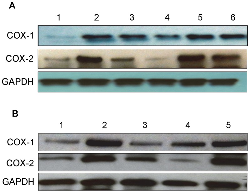
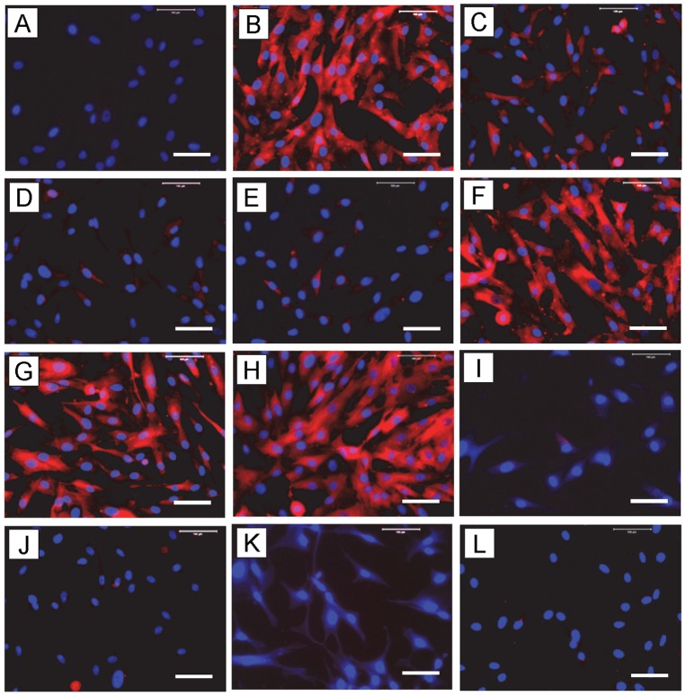
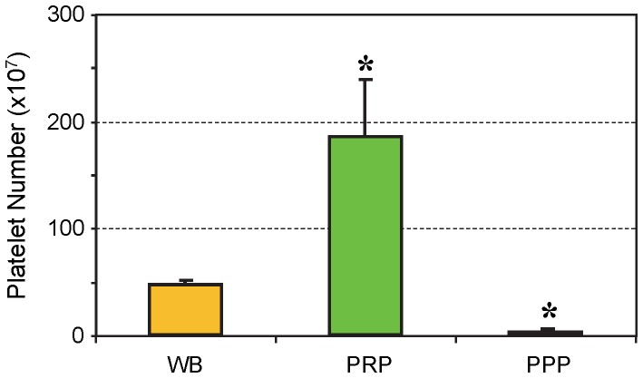
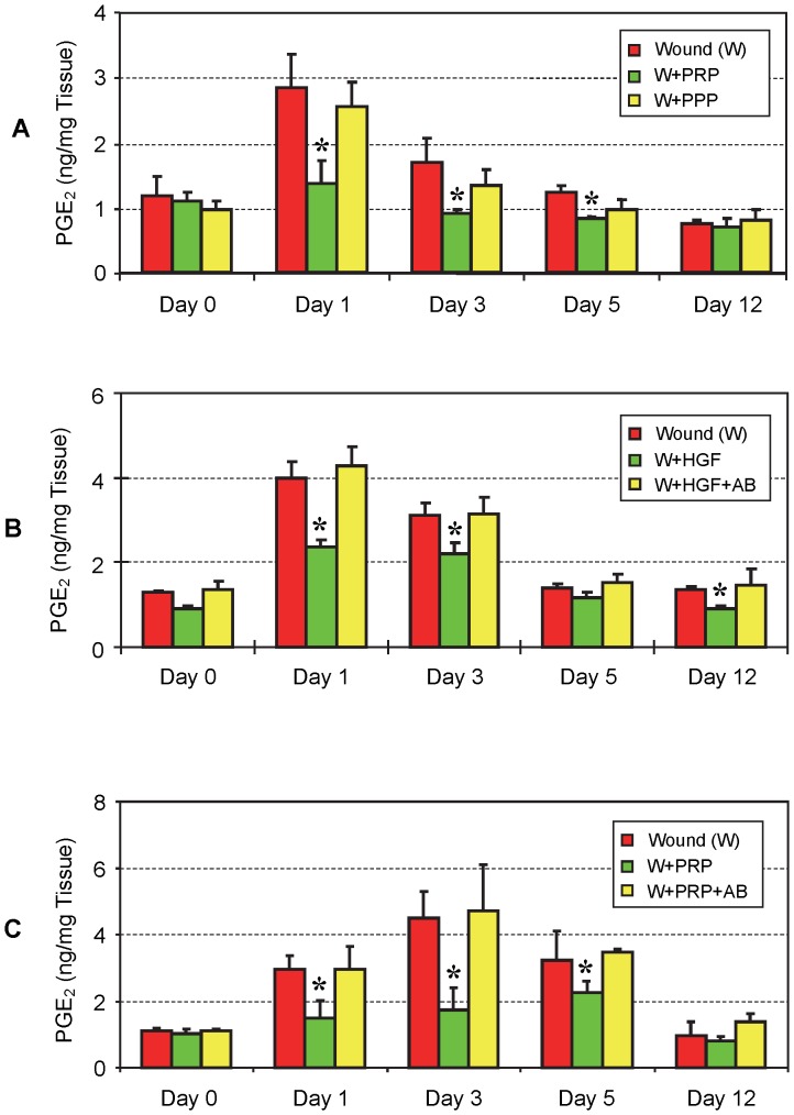
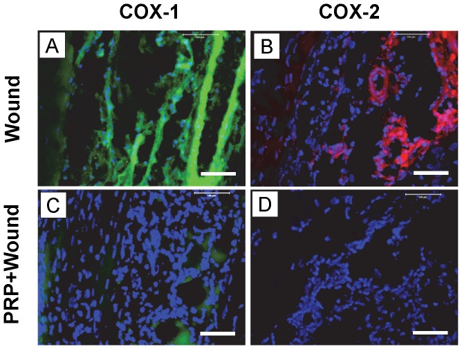
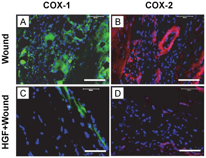
Platelet-rich plasma (PRP) containing hepatocyte growth factor (HGF) and other growth factors are widely used in orthopaedic/sports medicine to repair injured tendons. While PRP treatment is reported to decrease pain in patients with tendon injury, the mechanism of this effect is not clear. Tendon pain is often associated with tendon inflammation, and HGF is known to protect tissues from inflammatory damages. Therefore, we hypothesized that HGF in PRP causes the anti-inflammatory effects. To test this hypothesis, we performed in vitro experiments on rabbit tendon cells and in vivo experiments on a mouse Achilles tendon injury model. We found that addition of PRP or HGF decreased gene expression of COX-1, COX-2, and mPGES-1, induced by the treatment of tendon cells in vitro with IL-1β. Further, the treatment of tendon cell cultures with HGF antibodies reduced the suppressive effects of PRP or HGF on IL-1β-induced COX-1, COX-2, and mPGES-1 gene expressions. Treatment with PRP or HGF almost completely blocked the cellular production of PGE2 and the expression of COX proteins. Finally, injection of PRP or HGF into wounded mouse Achilles tendons in vivo decreased PGE2 production in the tendinous tissues. Injection of platelet-poor plasma (PPP) however, did not reduce PGE2 levels in the wounded tendons, but the injection of HGF antibody inhibited the effects of PRP and HGF. Further, injection of PRP or HGF also decreased COX-1 and COX-2 proteins. These results indicate that PRP exerts anti-inflammatory effects on injured tendons through HGF. This study provides basic scientific evidence to support the use of PRP to treat injured tendons because PRP can reduce inflammation and thereby reduce the associated pain caused by high levels of PGE2.


Conflict of interest statement

Competing Interests: The authors declare that they have no competing interests.

Figures

Figure 1
Figure 1. HGF levels in rabbit whole blood and PRP preparations.
The concentration of HGF in PRP was over 3.5 times the concentration found in rabbit whole blood (WB). The data are expressed as mean ± SD, n  = 7.
Figure 2
Figure 2. The effect of PRP (A) and HGF (B) treatment on COX-1 expression in tendon cell culture.
Tendon cells were treated with IL-1β, which resulted in high levels of COX-1 expression; however, the addition of PRP (A) or HGF (B) together with IL-1β to the cell culture markedly reduced the level of COX-1 expression. Also, PRP or HGF alone did not alter COX-1 expression. Note that *P<0.05 is with respect to IL-1β treatment (I); and #P<0.05 is with respect to the PRP+IL-1β (P+I) group in A and the HGF+IL-1β (H+I) group in B. The data are expressed as mean ± SD, n  = 4. SF: serum free medium; I: IL-1β; P+I: PRP+IL-1β; P+AB+I: PRP+HGF antibody+IL-1β; P: PRP; P+AB: PRP+HGF antibody; H+I: HGF+ IL-1β; H+AB+I: HGF+HGF antibody+IL-1β, H: HGF; and H+AB: HGF+HGF antibody. Concentrations of the reagents used were: IL-1β, 1 ng/ml; PRP, 10% (v/v); HGF, 1 ng/ml; HGF antibody, 10 ng/ml. For SD values that are barely visible, the variation in gene expression values was less than 5 (control group, SF is 1).
Figure 3
Figure 3. The effects of PRP (A) and HGF (B) treatment on COX-2 gene expression in tendon cell culture.
Both PRP and HGF treatments nearly abolished COX-2 expression, which was induced by IL-1β treatment of tendon cells. Note that *P<0.05 is with respect to IL-1β treatment condition (I); and #P<0.05 is with respect to the PRP+IL-1β (P+I) group in A and the HGF+IL-1β (H+I) group in B. The data are expressed as mean ± SD, n  = 4. Label abbreviations and concentrations of all agents (IL-1β, PRP, HGF, and HGF antibody) are identical to legends in Figure 2 . For SD values that are barely visible, the variation in gene expression values was less than 5 (control group, SF is 1).
Figure 4
Figure 4. The effects of PRP (A) and HGF (B) treatment on mPGES-1 gene expression in tendon cell culture.
IL-1β treatment increased mPGES-1 expression (I), but its effect was almost completely suppressed by PRP (A) or HGF (B) treatment. Note that *P<0.05 is with respect to IL-1β treatment condition (I); and #P<0.05 is with respect to the PRP+IL-1β (P+I) group in A and the HGF+IL-1β (H+I) group in B. The data are expressed as mean ± SD, n  = 4. Label abbreviations and concentrations of all agents (IL-1β, PRP, HGF, and HGF antibody) are identical to legends in Figure 2 . For SD values that are barely visible, the variation in gene expression values was less than 5 (control group, SF is 1).
Figure 5
Figure 5. The effects of PRP (A) and HGF (B) treatments on PGE2 production in tendon cell culture.
IL-1β treatment (I) induced high levels of PGE2 production in cells compared to levels induced by control conditions (SF); however, these increases in PGE2 were markedly reduced by PRP or HGF treatment. Note that *P<0.05 is with respect to IL-1β treatment condition (I); and #P<0.05 is with respect to the PRP+IL-1β (P+I) group in A and the HGF+IL-1β (H+I) group in B. The data are expressed as mean ± SD, n  = 4. Label abbreviations and concentrations of all agents (IL-1β, PRP, HGF, and HGF antibody) are identical to legends in Figure 2 . For SD values that are barely visible, the variation in gene expression values was less than 5 (control group, SF is 1).
Figure 6
Figure 6. The effects of PRP (A) and HGF (B) treatment on COX-1 and COX-2 protein expression in tendon cell culture.
Total proteins extracted from tendon cells after treatments were separated on 12% SDS-PAGE and transferred onto nitrocellulose membrane to detect COX-1 and COX-2 proteins using goat anti-COX-1 and goat anti-COX-2 polyclonal antibodies, respectively, and peroxidase-conjugated rabbit anti-goat IgG antibody as secondary antibody. A. PRP treatment. Lane 1: serum free medium (SF); lane 2: IL-1β (1 ng/ml); lane 3: PRP (2%)+IL-1β (1 ng/ml); lane 4: PRP (10%)+ IL-1β (1 ng/ml); lane 5: PRP (2%)+ IL-1β (1 ng/ml)+HGF antibody (10 ng/ml); lane 6: PRP (10%)+IL-1β (1 ng/ml)+HGF antibody (10 ng/ml). Notice that 10% PRP reduced COX-1 expression, but almost completely inhibited COX-2 expression (lane 4 vs. lane 2). B. HGF treatment. Lane 1: SF; lane 2: IL-1β (1 ng/ml); lane 3: HGF (1 ng/ml)+IL-1β (1 ng/ml); lane 4: HGF (1 ng/ml); and lane 5: HGF (1 ng/ml)+IL-1β (1 ng/ml)+HGF antibody (10 ng/ml). Also notice that HGF at 1 ng/ml markedly reduced both COX-1 and COX-2 expression (lane 3 vs. lane 2).
Figure 7
Figure 7. The effects of PRP or HGF treatment on COX-1 protein expression in tendon cell culture by immunostaining.
Tendon cells in culture were treated with IL-1β, PRP, HGF, and/or HGF antibody (AB) to determine COX-1 expression using goat anti-COX-1 antibody and Cy-3 conjugated donkey anti-goat IgG antibody. Red represents COX-1 protein; and blue represents nuclei stained with Hoechst 33342. IL-1β treatment induced a high level of COX-1 expression in tendon cells compared to the control conditions (A); however, such an increase in COX-1 was markedly reduced by PRP or HGF treatments. The addition of HGF antibody to tendon cell cultures decreased the reduction by PRP or HGF treatments. A: SF (control conditions); B: IL-1β (1 ng/ml); C: 2%PRP+ IL-1β; D: 10%PRP+IL-1β; E: HGF+IL-1β; F: 2%PRP+IL-1β+AB; G: 10%PRP+IL-1β+AB; H: HGF+IL-1β+AB; I: 2%PRP; J: 10%PRP; K: HGF (1 ng/ml) and L: HGF+AB (10 ng/ml). Bar: 100 µm.
Figure 8
Figure 8. The effects of PRP or HGF treatment on COX-2 protein expression in tendon cell culture by immunostaining.
Tendon cells in culture were treated with IL-1β, PRP, HGF, and/or HGF antibody (AB) to determine COX-2 expression using goat anti-COX-2 antibody and Cy-3 conjugated donkey anti-goat IgG antibody. Red represents COX-2 protein; and blue represents nuclei stained with Hoechst 33342. Similar to COX-1 results ( Figure 7 ), PRP or HGF treatments markedly reduced COX-2 expression, and the combined use of HGF antibody with PRP or HGF decreased the reduction effects produced by either PRP or HGF treatment alone. Label abbreviations (A to L) are the same as in Figure 7 . Bar: 100 µm.
Figure 9
Figure 9. Platelet numbers in mouse whole blood, PRP, and PPP preparations used for the in vivo experiments.
On average, the number of platelets in PRP preparations was about four times higher than in mouse whole blood (WB), whereas the number was 60 times higher in PRP than in PPP preparations (*P<0.05 is with respect to WB). The data are expressed as mean ± SD, and n  = 8.
Figure 10
Figure 10. The effects of PRP, PPP, HGF and HGF antibody on PGE2 production in wounded mouse Achilles tendons in vivo.
A. PRP and PPP injections. Injections with PRP significantly reduced PGE2 levels in tendons on days 1, 3, and 5 post-treatment. However, on day 12, PRP’s effect on PGE2 production in tendon tissues was little, if any. On day 0, little difference was observed in PGE2 levels between PRP-treated and non-treated tendons. However, PPP treatment did not significantly decrease PGE2 levels in the tendon tissues at all time points post-treatment (P>0.05). However, a small suppression effect on days 1, 3, and 5 likely occurred, as PPP-treated tendons had consistently lower PGE2 levels than control tendons injected with saline. B. HGF injection. Injection with HGF decreased PGE2 production in wounded Achilles tendons, producing results similar to PRP injections except on day 12, when HGF injection still significantly decreased PGE2 production (*P<0.01 is with respect to control tendons given saline injections). Addition of HGF+HGF antibody (AB) largely negated the suppressive effect of HGF. C. PGE2 production in mouse tendons injected with PRP and HGF antibody. Injection of PRP along with HGF antibody into wounded Achilles tendons restored PGE2 levels to normal on all days 1, 3, 5 and 12. The data in Figure 10A, B, C are expressed as mean ± SD, and n  = 4 (4 mice).
Figure 11
Figure 11. Immunostaining of COX-1 and COX-2 on tissue sections of wounded mouse Achilles on day 3 after wounding and PRP treatment.
Dissected tissues were cut, fixed in 4% paraformaldehyde. For COX-1, the tissue sections were stained with goat anti-COX-1 antibody and FITC-conjugated rabbit anti-goat IgG antibody. For COX-2, the tissue sections were stained with goat anti-COX-2 antibody and Cy-3 conjugated donkey anti-goat IgG antibody. Green represents COX-1; red represents COX-2; and blue represents nuclei stained with Hoechst 33342. Both COX-1 (A) and COX-2 (B) were positively stained on the wound sites of the mouse tendons without PRP treatment. However, treatment with PRP, with its platelet concentration about 4 times over that of mouse whole blood ( Figure 9 ), nearly abolished COX-1 (C) and COX-2 (D) expressions. Bar: 100 µm.
Figure 12
Figure 12. Immunostaining of COX-1 and COX-2 on tissue sections of wounded mouse Achilles tendons on day 3 after wounding and HGF treatment.
Mouse Achilles tendons were dissected 3 days after wounding and with or without HGF treatment followed by mincing and fixing tissues in 4% paraformaldehyde. For COX-1 and COX-2 staining, the tissue sections were stained with goat anti-COX-1 and goat anti-COX-2 antibodies respectively, and detected using FITC-conjugated rabbit anti-goat IgG antibody (COX-1) or Cy-3 conjugated donkey anti-goat IgG antibody (COX-2) respectively. Green represents COX-1; red represents COX-2; and blue represents nuclei stained with Hoechst 33342. Both COX-1 (A) and COX-2 (B) were positively stained on the wounded area without HGF treatment. However, HGF treatment (3 ng in 10 µl saline) decreased COX-1 (C) and COX-2 (D) expression. Bar: 100 µm.

References

    1. Butler DL, Juncosa N, Dressler MR (2004) Functional efficacy of tendon repair processes. Annu Rev Biomed Eng 6: 303–329. - PubMed
    1. Smith WL (1989) The eicosanoids and their biochemical mechanisms of action. Biochem J 259: 315–324. - PMC - PubMed
    1. Williams TJ (1979) Prostaglandin E2, prostaglandin I2 and the vascular changes of inflammation. Br J Pharmacol 65: 517–524. - PMC - PubMed
    1. Ferreira SH, Nakamura M, de Abreu Castro MS (1978) The hyperalgesic effects of prostacyclin and prostaglandin E2. Prostaglandins 16: 31–37. - PubMed
    1. Kopp UC, Cicha MZ (1999) PGE2 increases substance P release from renal pelvic sensory nerves via activation of N-type calcium channels. Am J Physiol 276: R1241–1248. - PubMed

Publication types

MeSH terms