. 2013 Jun 28;8(6):e67608.
doi: 10.1371/journal.pone.0067608. Print 2013.

MOR is not enough: identification of novel mu-opioid receptor interacting proteins using traditional and modified membrane yeast two-hybrid screens

Affiliations

MOR is not enough: identification of novel mu-opioid receptor interacting proteins using traditional and modified membrane yeast two-hybrid screens

Jessica Petko et al. PLoS One. .

Abstract

The mu-opioid receptor (MOR) is the G-protein coupled receptor primarily responsible for mediating the analgesic and rewarding properties of opioid agonist drugs such as morphine, fentanyl, and heroin. We have utilized a combination of traditional and modified membrane yeast two-hybrid screening methods to identify a cohort of novel MOR interacting proteins (MORIPs). The interaction between the MOR and a subset of MORIPs was validated in pulldown, co-immunoprecipitation, and co-localization studies using HEK293 cells stably expressing the MOR as well as rodent brain. Additionally, a subset of MORIPs was found capable of interaction with the delta and kappa opioid receptors, suggesting that they may represent general opioid receptor interacting proteins (ORIPS). Expression of several MORIPs was altered in specific mouse brain regions after chronic treatment with morphine, suggesting that these proteins may play a role in response to opioid agonist drugs. Based on the known function of these newly identified MORIPs, the interactions forming the MOR signalplex are hypothesized to be important for MOR signaling and intracellular trafficking. Understanding the molecular complexity of MOR/MORIP interactions provides a conceptual framework for defining the cellular mechanisms of MOR signaling in brain and may be critical for determining the physiological basis of opioid tolerance and addiction.


Conflict of interest statement

Competing Interests: The authors have declared that no competing interests exist.

Figures

Figure 1
Figure 1. Mapping the MOR/MORIP interaction.
Candidate MORIPs identified in the traditional and MYTH Y2H screens (preys) were tested for interaction with each of the intracellular loops (IL) and carboxyl-terminus (C-tail) of the mu-opioid receptor (baits) in a directed Y2H assay. An empty bait vector (pACT) was used as a negative control. A positive interaction is indicated by the production of a blue yeast colony in the β-galactosidase assay.
Figure 2
Figure 2. Confirmation of MORIP/MOR-IL2 interactions.
GST pulldown assays were performed to interrogate the interaction between selected full-length MORIPs and the MOR-IL2 domain. In the top three panels, MORIP-GST fusion proteins were used to pull down the S-tagged MOR-IL2. In the bottom three panels, MOR-IL2-GST fusion proteins were used to pull down S-tagged MORIPs. Pulldown products were purified on glutathione beads, separated by SDS-PAGE, and probed on Western blots using HRP-conjugated anti-S-tag antibodies. S-tagged MORIPs or MOR-IL2 domains produced in bacteria are shown in lysate lanes (Ly), while uncoated glutathione sepharose beads (Beads) or GST-coated glutathione sepharose beads (GST) incubated with S-tagged proteins served as negative controls. PD indicates pull-down lanes.
Figure 3
Figure 3. Interaction of MORIPs with MOR, DOR, and KOR in mammalian cells.
(A) Amino acid sequence comparison between MOR-, DOR-, and KOR-IL2 domains. (B) Co-immunoprecipitation of full length MORIPs with MOR, DOR, or KOR. FLAG-tagged MOR, DOR, or KOR was immunoprecipitated from HEK-MOR, HEK-DOR, or HEK-KOR cells, respectively, using rabbit anti-FLAG antibodies. Mock immunoprecipitations were performed with Protein-G beads coated with non-specific rabbit IGG. Blots were probed with either anti-MORIP antibodies for the presence of endogenously expressed MORIPs (SIAH1, SIAH2 and WLS) or with anti-myc antibodies for transiently transfected myc-tagged MORIPs. A representative blot from each column was probed for mouse anti-FLAG to confirm immunoprecipitation of MOR, DOR, and KOR from these cell lines. Lysate lanes (L) contain 5% of the total protein compared to the mock (M) and immunoprecipitation (IP) lanes.
Figure 4
Figure 4. MORIP expression and interaction with MOR in mouse brain.
(A) Expression of MOR and MORIPs in mouse brain regions. Western blots containing lysates prepared from mouse cerebellum (C), hippocampus (H), midbrain (M), nucleus accumbens (N), prefrontal cortex (P), and striatum (S) were probed with anti-MORIP antibodies. (B) Co-immuniprecipitation. The MOR was immunoprecipitated from whole mouse brain lysates using rabbit anti-MOR antibodies. Immunocomplexes were probed for the presence of SIAH1, VAPA, or WLS using MORIP-specific antibodies. Lysate lanes (L) contain 5% of the total protein prepared from whole mouse brain lysate compared to the mock (M, rabbit IGG) and immunoprecipitation (IP) lanes.
Figure 5
Figure 5. MORIP expression in brain regions of morphine-treated mice.
Mice were treated for 96 hours with either morphine-containing (n = 5) or placebo (n = 4) pellets. Animals were sacrificed, brain regions dissected, and Western blots of selected MORIPs probed with MORIP-specific antibodies. Each panel contains a representative blot for a MORIP in the specified brain region (n = 4 blots/MORIP/brain region). Total protein was quantified by Ponceau stain of the blot prior to antibody probing. Bar graphs represent the average pixel density (as determined by imageJ) of four blots for each brain region normalized to total protein and placebo treatment. Data was analyzed using a two-sided Student’s t-test. Error is expressed as standard error of the mean. * indicates a statistically significant difference (p<.05) between sham and morphine treatment.
Figure 6
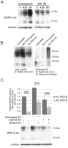
Figure 6. Regulation of MOR protein abundance and ubiquitination.
(A) Lysates (10 µg) from HEK-MOR cells treated with 200 µM chloroquine or 30 µM MG132 for 0, 4.5, or 9 hours were probed with anti-FLAG antibodies. All blots were normalized to GAPDH. (B) Lysates from HEK-MOR cells treated for 9 hours with the indicated inhibitors were immunoprecipitated with rabbit anti-FLAG and blots probed for mouse anti-ubiquitin (right panel). The amount of lysate loaded into each IP reaction was normalized to the amount of MOR detected in 10 µg of cell lysate (left panel) to ensure that equal amounts of MOR were being immunoprecipitated. Experiments for A and B were performed in triplicate. (C) HEK-MOR cells were treated for 6 hours with no proteolytic inhibitors, chloroquine, or MG132 in the presence or absence of 10 µM DADLE. Total cell lysates (10 µg) were probed for rabbit anti-FLAG. Bar graphs represent the average pixel density (as determined by ImageJ) from 5 experiments normalized to GAPDH and an untreated control (no inhibitor or DADLE). The average percent DADLE-induced decrease in MOR levels for each inhibitor treatment was compared to the percent reduction observed without inhibitor treatment using a two-sided paired Student’s t-test. Error is expressed as standard error of the mean. * (p<.01) and ** (p<.005) indicate statistical difference as compared to no inhibitor treatment. IP indicates the antibody used for immunoprecipitation while IB indicates the antibody used for immunoblotting.
Figure 7
Figure 7. Role of SIAH1 and SIAH2 in regulating MOR ubiquitination.
(A) Mapping the interaction site of MOR-IL2 on SIAH1 using GST-pulldowns. As controls, S-tagged constructs were incubated with untreated or GST-coated beads. (B) HEK-MOR cells were transfected with wild-type or truncated SIAH (trSIAH1 or trSIAH2) constructs and treated for 6 hours with 30 µM MG132. Proteins were immunoprecipitated and blots probed with either mouse anti-myc or mouse anti-HA to test for SIAH expression (left panels) and interaction with MOR (right panels), respectively. (C) HEK-MOR cells were transfected with wild-type or truncated SIAH constructs and either left untreated or treated with 10 µM DADLE for 6 hours. Blots were cut into sections and probed with rabbit anti-FLAG, mouse anti-myc, mouse anti-HA, or chicken anti-GAPDH antibodies. Bar graphs represent the average pixel density from 4 experiments normalized to GAPDH and untreated controls and subjected to a two-sided paired Student’s t-test. None of the SIAH constructs caused significant changes (at p<.05) in steady-state levels of MOR protein expression or in DADLE-mediated decreases in MOR expression levels. (D–F) Ubiquitination of MOR. Equal amounts of MOR (normalized from lysate blot) were loaded into immunoprecipitation reactions with anti-FLAG antibody. In each panel, the upper blot shows the IP probed for ubiquitin. All other blots show expression of various constructs or GAPDH in transfected cells. All experiments were performed in triplicate. (D) Steady-state ubiquitination levels in transfected HEK-MOR cells (E) Ubiquitination levels in transfected cells treated with 30 µM MG132 (F) Agonist induced ubiquitination in transfected cells treated with 10 µM DADLE.
Figure 8
Figure 8. Functional categories of known and novel MORIPS.
Newly identified and known MOR interacting proteins based on data from this and previous studies , , , , , , , , . MORIPs were grouped based on established functional properties. MORIPs identified in the current study are depicted by green shaded circles, while previously identified MORIPs are represented by white circles. Many of the newly identified MORIPs also interact with DOR and KOR and may be considered general ORIPS (see Results).

References

    1. Matthes HW, Maldonado R, Simonin F, Valverde O, Slowe S, et al. (1996) Loss of morphine-induced analgesia, reward effect and withdrawal symptoms in mice lacking the mu-opioid-receptor gene. Nature 383: 819–823. - PubMed
    1. Raehal KM, Bohn LM (2005) Mu opioid receptor regulation and opiate responsiveness. Aaps J 7: E587–591. - PMC - PubMed
    1. von Zastrow M, Svingos A, Haberstock-Debic H, Evans C (2003) Regulated endocytosis of opioid receptors: cellular mechanisms and proposed roles in physiological adaptation to opiate drugs. Curr Opin Neurobiol 13: 348–353. - PubMed
    1. Koch T, Hollt V (2008) Role of receptor internalization in opioid tolerance and dependence. Pharmacol Ther 117: 199–206. - PubMed
    1. Koch T, Widera A, Bartzsch K, Schulz S, Brandenburg LO, et al. (2005) Receptor endocytosis counteracts the development of opioid tolerance. Mol Pharmacol 67: 280–287. - PubMed

Publication types

MeSH terms