. 2013 Jun 28;8(6):e67961.
doi: 10.1371/journal.pone.0067961. Print 2013.

Hsp70 oligomerization is mediated by an interaction between the interdomain linker and the substrate-binding domain

Affiliations

Hsp70 oligomerization is mediated by an interaction between the interdomain linker and the substrate-binding domain

Francesco A Aprile et al. PLoS One. .

Abstract

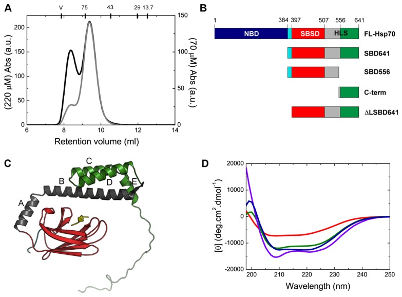
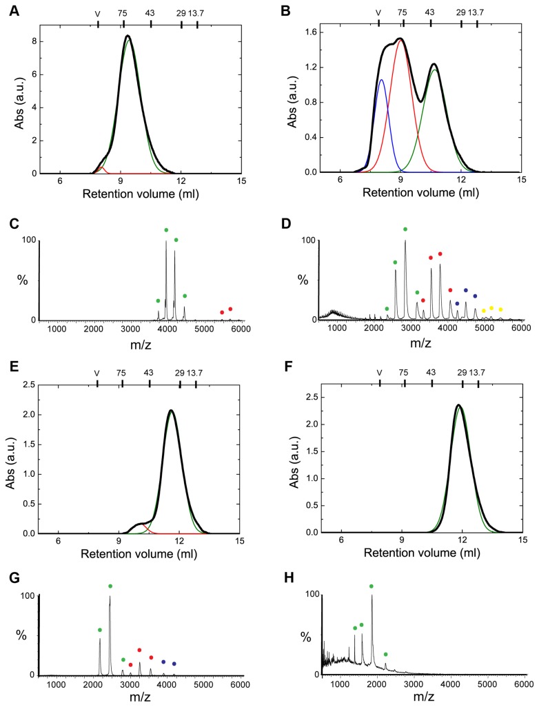
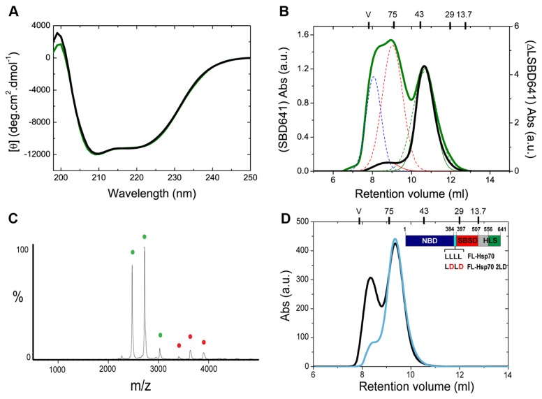
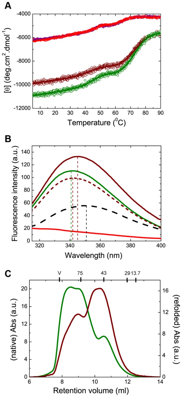
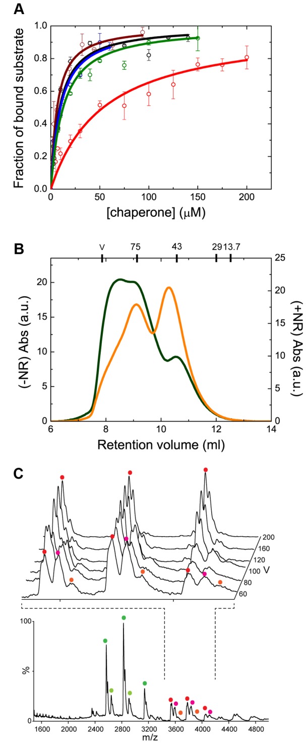
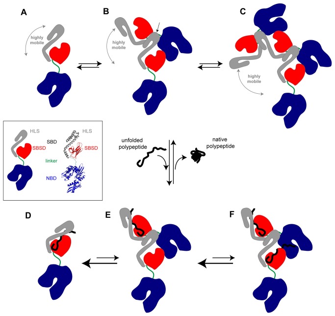
Oligomerization in the heat shock protein (Hsp) 70 family has been extensively documented both in vitro and in vivo, although the mechanism, the identity of the specific protein regions involved and the physiological relevance of this process are still unclear. We have studied the oligomeric properties of a series of human Hsp70 variants by means of nanoelectrospray ionization mass spectrometry, optical spectroscopy and quantitative size exclusion chromatography. Our results show that Hsp70 oligomerization takes place through a specific interaction between the interdomain linker of one molecule and the substrate-binding domain of a different molecule, generating dimers and higher-order oligomers. We have found that substrate binding shifts the oligomerization equilibrium towards the accumulation of functional monomeric protein, probably by sequestering the helical lid sub-domain needed to stabilize the chaperone: substrate complex. Taken together, these findings suggest a possible role of chaperone oligomerization as a mechanism for regulating the availability of the active monomeric form of the chaperone and for the control of substrate binding and release.


Conflict of interest statement

Competing Interests: The authors have declared that no competing interests exist.

Figures

Figure 1
Figure 1. The different human Hsp70 variants maintain native-like structure.
(A) Analytical SEC analysis of FL-Hsp70 at protein concentrations of 220 µM (black line) and 70 µM (grey line). At the top of the chromatogram the molecular weights of the following standard proteins used to calibrate the column are reported at the position which each elutes: conalbumin (75 kDa), ovalbumin (43 kDa), carbonic anhydrase (29 kDa) and ribonuclease A (13.7 kDa). The void volume (V) of the column (7.8 ml) was determined using blue dextran. (B) Schematic representation of the engineered human Hsp70 variants used in this study, showing the different functional domains for each variant: the nucleotide-binding domain, NBD, shown in dark blue, the interdomain linker shown in light blue and the substrate binding domain, SBD, which is composed of the substrate-binding subdomain, SBSD, shown in red, and the helical lid subdomain, HLS, with helices A-B shown in grey and helices C-E and the C-terminal tail shown in green (see text for a detailed description of the truncated variants). (C) Representation of the different truncated constructs on the crystal structure of the DnaK SBD in complex with the NR peptide substrate, represented in yellow (adapted from PDB ID: 1DKX [8]). (D) Far-UV CD spectra of the native FL-Hsp70 (blue), SBD641 (green), SBD556 (red) and C-term (violet) variants (see Table S1 in File S1 for a detailed analysis of the secondary structure content of each chaperone variant).
Figure 2
Figure 2. The different protein variants have different propensities to oligomerize.
Analytical SEC and nESI mass spectra of the different protein constructs measured at the same protein concentration (6 µM): FL-Hsp70 (A, C), SBD641 (B, D), SBD556 (E, G) and C-term (F, H) variants. The SEC elution profiles were fitted to multi-peak Gaussian functions to evaluate the relative fractions of monomeric and oligomeric protein species. The molecular masses of the standard proteins used to calibrate the SEC column are reported at the top of the chromatogram. The elution peaks in SEC and the peaks in MS (Table S2 in File S1) corresponding to monomeric protein are coloured in green for all the variants, while those for dimers are in red, trimers in blue and tetramers in yellow. Note that comparing visually the SEC and MS data quantitatively is difficult as the relative peak heights in the mass spectra report on molecular abundance, which is independent of oligomeric state, while the SEC data depend on molecular absorbance.
Figure 3
Figure 3. The interdomain linker is essential for chaperone oligomerization.
(A) Far-UV CD spectrum of native ∆LSBD641 (black) compared to that of the SBD641 variant (green). (B) Comparison of the SEC chromatograms obtained for SBD641 (in green) and ΔLSBD641 (in black) at a protein concentration of 6 µM. (C) nESI MS analysis of ΔLSBD641 (monomer in green, dimer in red). (D) Comparison of the SEC chromatograms obtained for FL-Hsp70 (in black) and FL-Hsp70 2LD (in cyan) at a protein concentration of 220 µM. The molecular masses of the standard proteins are again reported at the top of the chromatogram.
Figure 4
Figure 4. Misfolding of HLS prevents oligomerization.
(A) Far-UV CD data for the thermally-induced denaturation of the native and refolded protein SBD556 (red and purple circles, respectively) and SBD641 (green and brown, respectively); a three-state denaturation model was used to analyse the experimental data (continuous lines; see Materials and Methods and Table S3 in File S1). (B) Tryptophan (W580) fluorescence spectra of SBD641 in their native (green line), thermally denatured (black dashed line) and refolded (brown continuous line) states; the spectrum of SBD556 in its native state is shown as a negative control (red continuous line). The calculated spectrum of SBD641, corresponding to 80% of the protein molecules in the native structure and 20% of the molecules in the thermally denatured state, assuming that the total fluorescence signal at a given wavelength is a linear combination of the fluorescence signal for each populated state (brown dashed lines), is also shown for comparison with the spectrum of the protein in its refolded state. (C) Analytical SEC performed on native and refolded (after thermal denaturation of the protein) states of SBD641 (green and brown lines, respectively). The molecular weights of the standard proteins used to calibrate the column are reported at the top of the chromatogram.
Figure 5
Figure 5. Substrate binding affects the oligomerization equilibrium.
(A) Fluorescence titration of the binding of the NR peptide to the different Hsp70 constructs (showing the fraction of bound substrate as a function of chaperone concentration): SBD556 (red circles), native (green circles) and refolded (brown circles) SBD641, ΔLSBD641 (black circles), and FL-Hsp70 (blue circles). The continuous lines represent the best fits of the data to a single binding site model. (B) SEC analysis of SBD641 in the absence (green line) and the presence (orange line) of a 14-fold excess of NR peptide. (C) nESI MS spectrum of SBD641 in the presence of the NR peptide at a protein:peptide ratio of 1:1 at a sample cone voltage of 60 V. Inset is the region of the spectrum showing dimers, at increasing sample cone voltages from front to back. The coloured circles represent charge states that correspond to the same protein species: uncomplexed monomeric protein in dark green, monomeric SBD641 complexed with the NR peptide in light green, the uncomplexed dimeric protein in red, dimeric SBD641 complexed with a single NR peptide molecule in pink, and dimeric SBD641 complexed with two NR peptide molecules in orange. The protein: peptide molar ratio used allowed sufficient resolution of the various charge state series for unambiguous assignment of substrate-bound chaperone states, which are validated by the successive removal of peptide equivalents upon increasing collisional activation (inset). The spectra shown are representative of five repeats, each incorporating >150 scans, with the relative abundances of apo- and holo-forms varying by <5% between individual nanoESI injections. Observed masses differ from those expected, due to residual adduction of solvent molecules and buffer ions, by <1%, as is typical in the spectra of non-covalent complexes (Table S6 in File S1) [57].
Figure 6
Figure 6. Model for Hsp70 oligomerization.
Hsp70 is composed of a nucleotide-binding domain (NBD) and a substrate-binding domain (SBD), which are connected to each other by a highly conserved hydrophobic linker (see insert). The SBD is in turn divided into two subdomains: the substrate-binding subdomain (SBSD), with mostly β-sheet structure, and the helical lid subdomain (HLS) that is rich in α-helical structure. The schematic images of the Hsp70 molecule are based on the crystal structure of DnaK complexed with ADP and substrate (pdb: 2KHO [48]). In the absence of nucleotide and of a polypeptide substrate, Hsp70 exists at equilibrium as a distribution of monomers (state A), dimers (state B), and higher order oligomers (state C). In the monomeric state, the two functional domains are allosterically independent and the HLS of the SBD is highly mobile. Our results show that chaperone oligomerization occurs through an interaction between the HLS of one protein molecule and the interdomain linker of another molecule (this interaction is shown by an arrow in state B), allowing the formation of oligomeric species. In the presence of an unfolded polypeptide substrate, however, the equilibrium between the different states of the chaperone is shifted towards the accumulation of monomeric protein, which is the state that shows the highest affinity for substrates because of the stabilization of the complex by the HLS that acts as a lid preventing substrate release (state D). The oligomers, however, have only one HLS in each species that is free from interactions with the linker of another protein molecule, and are therefore able to stabilize the binding of only one substrate molecule; this protein molecule will, however, be able to stabilize the bound substrate (state E). The remaining HLSs interact with another molecule of the chaperone, and therefore they are unable to act as a lid to their corresponding substrate-binding pockets, resulting in a decreased affinity for substrate (state F). As a consequence of this fact, the overall affinity for the substrate per chaperone molecule is decreased in the oligomeric species when compared with the monomeric protein. Indeed, the higher the order of the oligomer, the higher the number of low-affinity binding pockets per chaperone molecule and the lower the overall affinity of the chaperone molecule for the substrate. Once the substrate folds (to give the native state), its hydrophobic residues are no longer able to interact with the binding pocket of the chaperone, which results in substrate release and the chaperone recovers its initial state (an equilibrium between states A, B and C).

References

    1. Evans CG, Chang L, Gestwicki JE (2010) Heat shock protein 70 (hsp70) as an emerging drug target. J Med Chem 53: 4585-4602. doi:10.1021/jm100054f. PubMed: 20334364. - DOI - PMC - PubMed
    1. Gupta RS, Singh B (1994) Phylogenetic analysis of 70 kD heat shock protein sequences suggests a chimeric origin for the eukaryotic cell nucleus. Curr Biol 4: 1104-1114. doi:10.1016/S0960-9822(00)00249-9. PubMed: 7704574. - DOI - PubMed
    1. Mayer MP, Schröder H, Rüdiger S, Paal K, Laufen T et al. (2000) Multistep mechanism of substrate binding determines chaperone activity of Hsp70. Nat Struct Biol 7: 586-593. - PubMed
    1. Vogel M, Mayer MP, Bukau B (2006) Allosteric regulation of Hsp70 chaperones involves a conserved interdomain linker. J Biol Chem 281: 38705-38711. doi:10.1074/jbc.M609020200. PubMed: 17052976. - DOI - PubMed
    1. Liu Q, Hendrickson WA (2007) Insights into Hsp70 chaperone activity from a crystal structure of the yeast Hsp110 Sse1. Cell 131: 106-120. doi:10.1016/j.cell.2007.08.039. PubMed: 17923091. - DOI - PMC - PubMed

Publication types

MeSH terms

Substances