. 2013 Jul 1;8(7):e68574.
doi: 10.1371/journal.pone.0068574. Print 2013.

Recombinant human adenovirus-p53 injection induced apoptosis in hepatocellular carcinoma cell lines mediated by p53-Fbxw7 pathway, which controls c-Myc and cyclin E

Affiliations

Recombinant human adenovirus-p53 injection induced apoptosis in hepatocellular carcinoma cell lines mediated by p53-Fbxw7 pathway, which controls c-Myc and cyclin E

Kangsheng Tu et al. PLoS One. .

Retraction in

Abstract

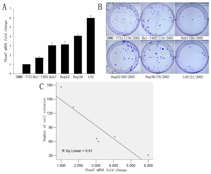
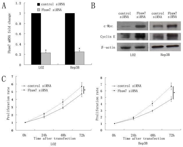
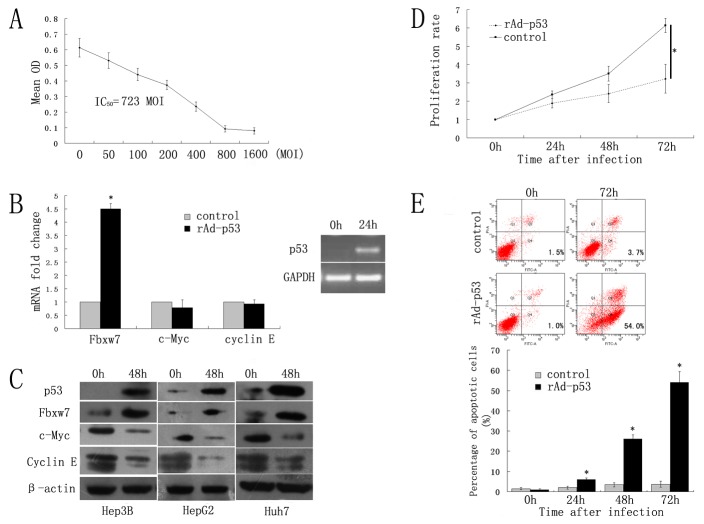
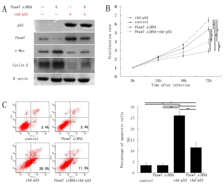
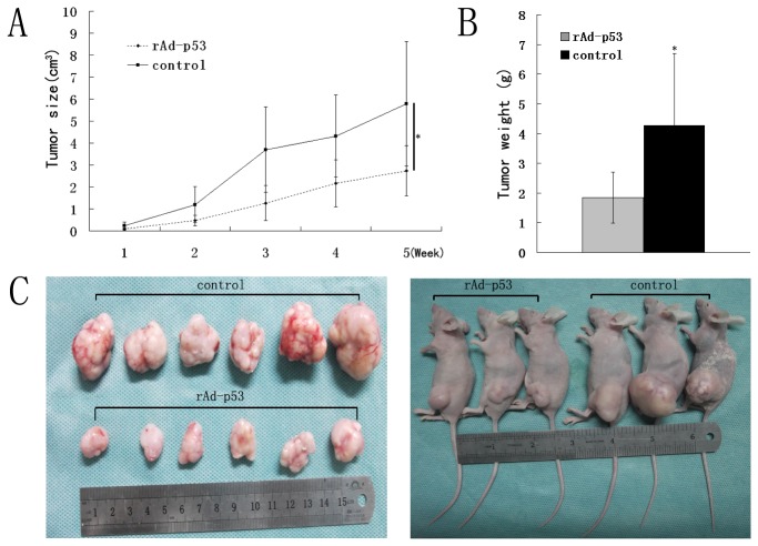
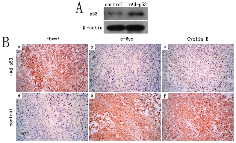
F-box and WD repeat domain-containing 7 (Fbxw7/hAgo/hCdc4/Fbw7) is a p53-dependent tumor suppressor and leads to ubiquitination-mediated suppression of several oncoproteins including c-Myc, cyclin E, Notch, c-Jun and others. Our previous study has indicated that low expression of Fbxw7 was negatively correlated with c-Myc, cyclin E and mutant-p53 in hepatocellular carcinoma (HCC) tissues. But the role and mechanisms of Fbxw7 in HCC are still unknown. Here, we investigated the function of Fbxw7 in HCC cell lines and the anti-tumor activity of recombinant human adenovirus-p53 injection (rAd-p53, Gendicine) administration in vitro and in vivo. Fbxw7-specific siRNA enhanced expression of c-Myc and cyclin E proteins and increased proliferation in cell culture. rAd-p53 inhibited tumor cell growth with Fbxw7 upregulation and c-Myc and cyclin E downregulation in vitro and a murine HCC model. This effect could be partially reverted using Fbxw7-specific siRNA. Here, we suggest that the activation of Fbxw7 by adenoviral delivery of p53 leads to increased proteasomal degradation of c-Myc and cyclin E enabling growth arrest and apoptosis. Addressing this pathway, we identified that rAd-p53 could be a potential therapeutic agent for HCC.


Conflict of interest statement

Competing Interests: The authors have declared that no competing interests exist.

Figures

Figure 1
Figure 1. Fbxw7 mRNA expression correlates negatively colony formation ability in HCC cell lines.
A) The Fbxw7 mRNA expression level of HCC cell lines (n=3). B) Colony formation assay quantification with HCC cell lines. C) Correlation plot for Fbxw7 mRNA levels and colony number. Values are depicted as mean±standard error.
Figure 2
Figure 2. Effect of Fbxw7 knockdown on LO2 and Hep3B cell lines.
A) qRT-PCR confirmed knockdown in LO2 and Hep3B cells treated with Fbxw7 siRNA. B) Expression of c-Myc and Cyclin E proteins were enhanced by Fbxw7 siRNA, as confirmed by Western blot analysis. β-actin served as loading control. C) Proliferation rates were assessed by MTT assay. Cell viability was normalized to time point 0 h. The proliferation rate of LO2 and Hep3B cells treated with Fbxw7 siRNA was significantly greater than in control siRNA cells. Values are depict as mean±standard error, n=3. *P<0.05 vs control siRNA.
Figure 3
Figure 3. rAd-p53 induces apoptosis in HCC cell lines with increased Fbxw7 and decreased c-Myc and Cyclin E.
A) MTT activity was measured after infecting Hep3B cells with rAd-p53 in an MOI gradient to determine their IC50 after 48 h. Mean OD values were used to calculate the IC50 via the modified Kou-type method: lgIC50 = Xm-I (P-(3-Pm-Pn)/4), in which Xm: lg maximum dose; I: lg (maximum dose/ adjacent dose); P: sum of positive response rate; Pm: the largest positive response rate; Pn: the smallest positive response rate. B) Over-expression of p53 24 h after infection as confirmed by RT-PCR. Quantification of RT-PCR for p53 and Fbxw7 indicated elevated mRNA levels for rAd-p53 infected Hep3B cells, whereas c-Myc and Cyclin E levels were not significantly decreased. C) Protein expression of p53 and Fbxw7 was enhanced, while c-Myc and Cyclin E expression was suppressed by rAd-p53 in Hep3B, HepG2 and Huh7 cells, as confirmed by Western blot analysis. β-actin served as loading control. D) Hep3B proliferation rate assessed by MTT assay was significantly lower in rAd-p53 infected cells compared to control cells. E) Quantification of apoptotic cell population (AnnexinV positive/ PI negative) by flow cytometry. rAd-P53 infected Hep3B cells were composed of a larger subset of apoptotic cells after 72 hours of infection compared to control. Values are depicted as mean±standard error, n=3. *P<0.05 vs control.
Figure 4
Figure 4. rAd-p53 induced apoptosis was reverted by Fbxw7-specific siRNA.
A) Fbxw7-siRNA treatment in Hep3B successfully down-regulated Fbxw7 protein but enhanced c-Myc and Cyclin expression as shown by immunostaining. Over-expression of rAd-p53 in the same cell line could enhance the levels of Fbxw7 and decrease c-Myc and Cyclin E expression. Fbxw7 knockdown in p53 over-expressing cells rescued partially the phenotype showing lower Fbxw7 levels with higher c-Myc and Cyclin E expression. β-actin served as loading control. B) The proliferation rate of rAd-p53 infected Hep3B cells was increased in the presence of Fbxw7-specific siRNA (MTT assay). C) The percentage of apoptotic Hep3B cells infected with rAd-p53 was decreased by Fbxw7-specific siRNA. Values are mean±standard error, n=3. * P<0.05 vs control; ** P<0.05 vs Fbxw7 siRNA; *** P<0.05 vs rAd-p53.
Figure 5
Figure 5. rAd-p53 inhibits tumor growth in vivo.
Multicenter intratumoral administration of rAd-P53 in Hep3B subcutaneous tumor in nude mice was monitored over time. Tumor size and weight was plotted in A), and B) respectively. C) Exemplarily, tumor bearing mice and explanted tumors are depicted. Values are mean±standard error. * P<0.05 vs rAd-p53.
Figure 6
Figure 6. p53-Fbxw7 pathway involvement in rAd-p53 treated tumor bearing mice.
A) Immonostaining of tissue homogenates confirm p53 over-expression in rAd-p53 treated mice, n=3. β-actin served as loading control. B) Fbxw7, c-Myc and Cyclin E were detected by SP–IHC on paraffin sections of both groups. Tumors treated with rAd-p53 showed strong signal for Fbsw7 protein (a), while c-Myc and Cyclin E were not detectable in the same tissue section (b and c). In saline treated tumors, weak Fbxw7 staining (d) with strong c-Myc and Cyclin E staining (e and f) was detected. Micrographs were acquired at 400 fold magnification, n=6.

Similar articles

Cited by

References

    1. Forner Alejandro, Llovet Josep M, Bruix Jordi (2012) Hepatocellular carcinoma. Lancet 379: 1245-1255. doi:10.1016/S0140-6736(11)61347-0. PubMed: 22353262. - DOI - PubMed
    1. Wertz IE, Kusam S, Lam C, Okamoto T, Sandoval W et al. (2011) Sensitivity to antitubulin chemotherapeutics is regulated by MCL1 and FBW7. Nature 471: 110-114. doi:10.1038/nature09779. PubMed: 21368834. - DOI - PubMed
    1. Crusio KM, King B, Reavie LB, Aifantis I (2010) The ubiquitous nature of cancer: the role of the SCF (Fbw7) complex in development and transformation. Oncogene 29: 4865-4873. doi:10.1038/onc.2010.222. PubMed: 20543859. - DOI - PMC - PubMed
    1. Wang Z, Fukushima H, Gao D, Inuzuka H, Wan L et al. (2011) The two faces of FBW7 in cancer drug Resistance. Bioessays 33: 851-859. doi:10.1002/bies.201100101. PubMed: 22006825. - DOI - PMC - PubMed
    1. Tu K, Zheng X, Yin G, Zan X, Yao Y et al. (2012) Evaluation of Fbxw7 expression and its correlation with expression of SREBP-1 in a mouse model of NAFLD. Mol Med Report 6: 525-530 - PubMed

Publication types

MeSH terms