. 2013 Jul 1;8(7):e68665.
doi: 10.1371/journal.pone.0068665. Print 2013.

Human DDX3 interacts with the HIV-1 Tat protein to facilitate viral mRNA translation

Affiliations

Human DDX3 interacts with the HIV-1 Tat protein to facilitate viral mRNA translation

Ming-Chih Lai et al. PLoS One. .

Abstract

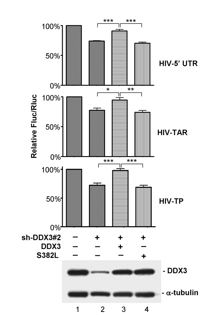
Nuclear export and translation of intron-containing viral mRNAs are required for HIV-1 gene expression and replication. In this report, we provide evidence to show that DDX3 regulates the translation of HIV-1 mRNAs. We found that knockdown of DDX3 expression effectively inhibited HIV-1 production. Translation of HIV-1 early regulatory proteins, Tat and rev, was impaired in DDX3-depleted cells. All HIV-1 transcripts share a highly structured 5' untranslated region (UTR) with inhibitory elements on translation of viral mRNAs, yet DDX3 promoted translation of reporter mRNAs containing the HIV-1 5' UTR, especially with the transactivation response (TAR) hairpin. Interestingly, DDX3 directly interacts with HIV-1 Tat, a well-characterized transcriptional activator bound to the TAR hairpin. HIV-1 Tat is partially targeted to cytoplasmic stress granules upon DDX3 overexpression or cell stress conditions, suggesting a potential role of Tat/DDX3 complex in translation. We further demonstrated that HIV-1 Tat remains associated with translating mRNAs and facilitates translation of mRNAs containing the HIV-1 5' UTR. Taken together, these findings indicate that DDX3 is recruited to the TAR hairpin by interaction with viral Tat to facilitate HIV-1 mRNA translation.


Conflict of interest statement

Competing Interests: HSS is an academic editor for PLOS ONE and this does not alter the adherence to all the PLOS ONE policies on sharing data and materials.

Figures

Figure 1
Figure 1. Knockdown of DDX3 by short hairpin RNAs inhibits HIV-1 production.
HeLa cells were mock-transfected with empty pSilencer 1.0-U6 vector (lane 1) or transfected with the pSilencer 1.0-U6 vector expressing a shRNA (lanes 2-5), respectively. After 36 hours, cells were re-transfected with HIV-1 proviral DNA pHXB2gpt plasmid, pEGFP-N1 and the same shRNA-expressing vectors. Transfected cells were cultured for another 48 hours, and then harvested for analysis. Immunoblotting was performed using antibodies against DDX3, eIF4A1, HIV-1 p24, GFP and α-tubulin. Viral production was assessed by detecting the expression levels of the HIV-1 Pr55Gag precursor and the mature CAp24 antigen. GFP and α-tubulin served as internal controls.
Figure 2
Figure 2. Translation of HIV-1 mRNAs is impaired in DDX3-depleted cells.
HeLa cells were transfected with empty pSilencer 1.0-U6 vector (mock) or the pSilencer 1.0-U6 vector expressing sh-DDX3#2 (DDX3-KD). After 72 hours, cells were re-transfected with the proviral DNA pHXB2gpt plasmid and harvested at 12 h post-transfection. A. Immunoblotting was performed using anti-DDX3 and anti-α-tubulin antibodies to show the knockdown efficiency of DDX3 in HeLa cells . B. Cytoplasmic extracts prepared from mock-transfected (mock) or DDX3-depleted (DDX3-KD) HeLa cells were subjected to 15-40% sucrose gradient sedimentation. RNA extracted from gradient fractions was analyzed by conventional RT-PCR using specific primers for HIV-1 Tat and Rev mRNAs (upper two panels). The housekeeping gene β-actin mRNA, whose translation is not significantly affected by DDX3 knockdown, served as a negative control (the 3rd panel). The translational efficiency of each mRNA was calculated as the ratio of polysome-associated mRNAs (fractions 7-11) to total mRNA (all fractions). The 18S and 28S rRNAs were resolved on a 1% formaldehyde/agarose gel and visualized by ethidium bromide staining (lower panel). C. RNA extracted from the cytoplasmic (Cyto.) and nuclear (Nu.) fractions of mock-transfected (mock) and DDX3-depleted (DDX3-KD) HeLa cells was analyzed by conventional RT-PCR using specific primers for HIV-1 Tat, HIV-1 Rev, and β-actin mRNAs (upper three panels). The subcellular fractions were also subjected to immunoblotting using anti-lamin A/C and anti-α-tubulin (lower two panels).
Figure 3
Figure 3. DDX3 facilitates translation of reporter mRNAs containing the 5’ UTR of HIV-1 mRNAs.
A. Schematic representation of the 5’ UTR of HIV-1 mRNAs. The 5’ UTR of all HIV-1 transcripts shares the same 289 nt noncoding region, which contains numerous cis-acting elements, including the transactivation response (TAR) hairpin, the polyadenylation (poly(A)) hairpin, the primer binding site (PBS), the dimerization initiation site (DIS), and the major splice donor site (SD). RNA secondary structures in the 5’ UTR of HIV-1 mRNA were adapted from Wilkinson et al. [24]. The functional motifs are indicated above each stem-loop domain. B. Schematic representation of the firefly luciferase (Fluc) reporters and details on the 5’ UTR of the pFL-SV40 derived reporters. HIV-5’ UTR represents the complete 5’ UTR of HIV-1 mRNAs (nucleotides 1-289). HIV-TAR contains the TAR hairpin (nucleotides 1-57), while HIV-5’ UTRΔTAR encompasses the 5’ UTR devoid of the TAR hairpin (nucleotides 58-289). HIV-TP contains the TAR-poly(A) region (nucleotides 1-104), while HIV-5’ UTRΔTP encompasses the 5’ UTR devoid of the TAR-poly(A) region (nucleotides 105-289). C. HeLa cells were co-transfected with a pFL-SV40 derived reporter and the control pRL-SV40 vector encoding the Renilla luciferase (Rluc) together with empty pSilencer 1.0-U6 vector (mock) or the pSilencer 1.0-U6 vector expressing sh-DDX3#2 (DDX3-KD) for 48 h. For each transfectant, the Fluc activity was first normalized to that of the Rluc control. Normalized Fluc activity of sh-DDX3#2-transfectants (DDX3-KD) was then compared to that of the mock. The bar graph shows the relative Fluc/Rluc activities in DDX3-KD cells relative to mock cells (upper panel). The pFL-SV40 derived reporter containing the 5’ UTR of cyclin E1 (CCNE1) served as a positive control. Fluc and Rluc mRNAs were analyzed by quantitative RT-PCR (lower panel). All data are shown as mean (± SEM) from at least three independent experiments (* p < 0.05, ** p < 0.01, *** p < 0.001).
Figure 4
Figure 4. The RNA helicase activity of DDX3 is required for translation of reporter mRNAs containing the HIV-1 5’ UTR.
HeLa cells were co-transfected with a pFL-SV40 derived reporter (HIV-5’ UTR, HIV-TAR or HIV-TP) and the control pRL-SV40 vector together with empty pSilencer 1.0-U6 vector (lane 1) or the pSilencer 1.0-U6 vector expressing sh-DDX3#2 (lanes 2-4) and a vector expressing shRNA-resistant wild-type DDX3 (lane 3) or S382L mutant (lane 4) for 48 h. For each transfectant, the Fluc activity was normalized to that of the Rluc control. The bar graph shows the relative Fluc/Rluc activities of each transfection relative to that of the mock transfection (lane 1). All data are shown as mean (± SEM) from at least three independent experiments (* p < 0.05, ** p < 0.01, *** p < 0.001). Immunoblotting was performed using anti-DDX3 and anti-α-tubulin, and representative data is shown (lower panel).
Figure 5
Figure 5. DDX3 interacts with HIV-1 Tat in vitro and in vivo.
A. His-tagged recombinant HIV-1 Tat protein was incubated with GST or GST-DDX3. After GST pull-down, bound proteins were analyzed by immunoblotting with anti-HIV-1 Tat antibody (upper panel). The GST-fusion proteins were resolved by SDS-PAGE and visualized by Coomassie blue staining (lower panel). B. The experiment was essentially similar to panel A, except that His-tagged recombinant HIV-1 Rev protein was used in the GST pull-down assay. Bound proteins were analyzed by immunoblotting with anti-HIV-1 Rev antibody (upper panel). C. The experiment was essentially similar to panel A. The assay used recombinant GST, GST-DDX3 (full-length; FL) or GST-DDX3 fragments (amino acids 1-226, 227-535 and 536-661) as bait to pull down His-tagged recombinant HIV-1 Tat protein. Bound proteins were analyzed by immunoblotting with anti-HIV-1 Tat antibody (upper panel). The GST-fusion proteins were resolved by SDS-PAGE and visualized by Coomassie blue staining (lower panel). D. FLAG-tagged DDX3 and HIV-1 Tat proteins were transiently co-expressed in HEK293 cells for 48 h. Immunoprecipitation was performed using anti-FLAG M2 agarose. Precipitated proteins were subjected to immunoblotting with anti-HIV-1 Tat antibody (upper panel) or anti-DDX3 antibody (lower panel). Ig H represents the immunoglobulin heavy chain. E. HIV-1 Tat protein was transiently expressed in HEK293 cells for 48 h. Immunoprecipitation was performed using anti-HIV-1 Tat antibody bound to protein A sepharose beads. Precipitated proteins were subjected to immunoblotting with anti-DDX3 antibody (upper panel) or anti-HIV-1 Tat antibody (lower panel).
Figure 6
Figure 6. HIV-1 Tat is co-localized with DDX3 in cytoplasmic stress granules under stress conditions.
A. GFP-DDX3 and HIV-1 Tat were transiently co-expressed in HeLa cells for 24 h. Immunofluorescent staining was carried out using mouse anti-HIV-1 Tat antibody. Subcellular localization of GFP-DDX3 (green) and HIV-1 Tat (red) was observed under a fluorescence microscope. The arrow indicates the co-localization of DDX3 and HIV-1 Tat in cytoplasmic foci. B. GFP-DDX3 was transiently expressed in HeLa cells for 24 h. Immunofluorescent staining was carried out using goat anti-TIA-1 antibody. Co-localization of GFP-DDX3 (green) and TIA-1 (red) in cytoplasmic SGs was observed under a fluorescence microscope. C. HeLa cells were transfected with a vector expressing HIV-1 Tat protein. At 24 h post-transfection, cells were mock-treated (mock) or treated with 0.5 mM sodium arsenite (arsenite) for 1 h. Immunofluorescent staining was carried out using rabbit anti-eIF4G and mouse anti-HIV-1 Tat antibodies. Subcellular localization of eIF4G (green) and HIV-1 Tat (red) was observed under a fluorescence microscope. The arrow indicates the co-localization of eIF4G and HIV-1 Tat in cytoplasmic SGs.
Figure 7
Figure 7. HIV-1 Tat is associated with translating mRNAs and facilitates translation of reporter mRNAs containing the HIV-1 5’ UTR.
A. HIV-1 Tat protein was transiently co-expressed with the Fluc reporter mRNA containing the 5’ UTR of HIV-1 mRNAs (HIV-5’ UTR) in HEK293 cells for 24 h. Cytoplasmic extracts weresubjectedto 15-40% sucrose gradient sedimentation. Proteins and RNAs were recovered from 23 fractions for analysis. Immunoblotting analysis of gradient fractions was performed using antibodies against DDX3, eIF4A1, eIF2α, eIF4E and HIV-1 Tat. The association of HIV-1 Tat with translation initiation complexes and polysomes was also detected in the absence of the HIV-1 5’ UTR reporter (w/o HIV-5’ UTR) and in DDX3-depleted (DDX3-KD) HEK293 cells. The 18S and 28S rRNAs were resolved on a 1% formaldehyde/agarose gel and visualized by ethidium bromide staining (lower panel). B. The in vitro translation assay was performed using in vitro-transcribed HIV-1 5’ UTR-containing Fluc mRNA and control Rluc mRNA as templates in HeLa cell lysate supplemented with different amounts of recombinant HIV-1 Tat. The graph shows the relative Fluc/Rluc activities of each reaction relative to that of the corresponding reaction without addition of HIV-1 Tat. All data are shown as mean (± SEM) from at least three independent experiments. C. The experiment was essentially similar to panel B, except that Rluc mRNA harbored the HIV-1 5’ UTR while the non-modefied Fluc mRNA served as a control. The graph shows the relative Rluc/Fluc activities of each reaction relative to that of the corresponding reaction without addition of HIV-1 Tat. All data are shown as mean (± SEM) from at least three independent experiments.

Similar articles

Cited by

References

    1. Chao CH, Chen CM, Cheng PL, Shih JW, Tsou AP et al. (2006) DDX3, a DEAD box RNA helicase with tumor growth-suppressive property and transcriptional regulation activity of the p21waf1/cip1 promoter, is a candidate tumor suppressor. Cancer Res 66: 6579-6588. doi:10.1158/0008-5472.CAN-05-2415. PubMed: 16818630. - DOI - PubMed
    1. Botlagunta M, Vesuna F, Mironchik Y, Raman A, Lisok A et al. (2008) Oncogenic role of DDX3 in breast cancer biogenesis. Oncogene 27: 3912-3922. doi:10.1038/onc.2008.33. PubMed: 18264132. - DOI - PMC - PubMed
    1. Kanai Y, Dohmae N, Hirokawa N (2004) Kinesin transports RNA: isolation and characterization of an RNA-transporting granule. Neuron 43: 513-525. doi:10.1016/j.neuron.2004.07.022. PubMed: 15312650. - DOI - PubMed
    1. Choi YJ, Lee SG (2012) The DEAD-box RNA helicase DDX3 interacts with DDX5, co-localizes with it in the cytoplasm during the G2/M phase of the cycle, and affects its shuttling during mRNP export. J Cell Biochem 113: 985-996. doi:10.1002/jcb.23428. PubMed: 22034099. - DOI - PubMed
    1. Shih JW, Tsai TY, Chao CH, Wu Lee YH (2008) Candidate tumor suppressor DDX3 RNA helicase specifically represses cap-dependent translation by acting as an eIF4E inhibitory protein. Oncogene 27: 700-714. doi:10.1038/sj.onc.1210687. PubMed: 17667941. - DOI - PubMed

Publication types