. 2013 Jul 1;8(7):e69042.
doi: 10.1371/journal.pone.0069042. Print 2013.

Enteric glia cells attenuate cytomix-induced intestinal epithelial barrier breakdown

Affiliations

Enteric glia cells attenuate cytomix-induced intestinal epithelial barrier breakdown

Gerald A Cheadle et al. PLoS One. .

Abstract

Background: Intestinal barrier failure may lead to systemic inflammation and distant organ injury in patients following severe injury. Enteric glia cells (EGCs) have been shown to play an important role in maintaining gut barrier integrity through secretion of S-Nitrosoglutathione (GSNO). We have recently shown than Vagal Nerve Stimulation (VNS) increases EGC activation, which was associated with improved gut barrier integrity. Thus, we sought to further study the mechanism by which EGCs prevent intestinal barrier breakdown utilizing an in vitro model. We postulated that EGCs, through the secretion of GSNO, would improve intestinal barrier function through improved expression and localization of intestinal tight junction proteins.

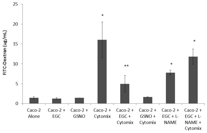
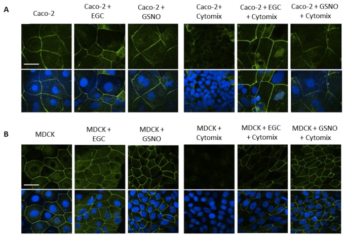
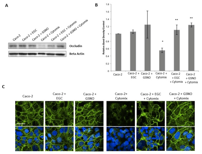
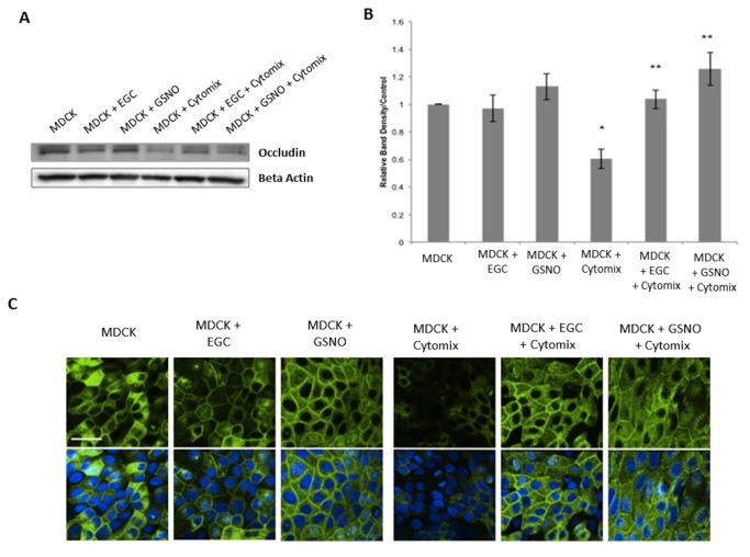
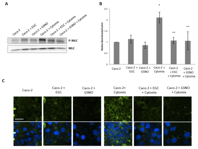
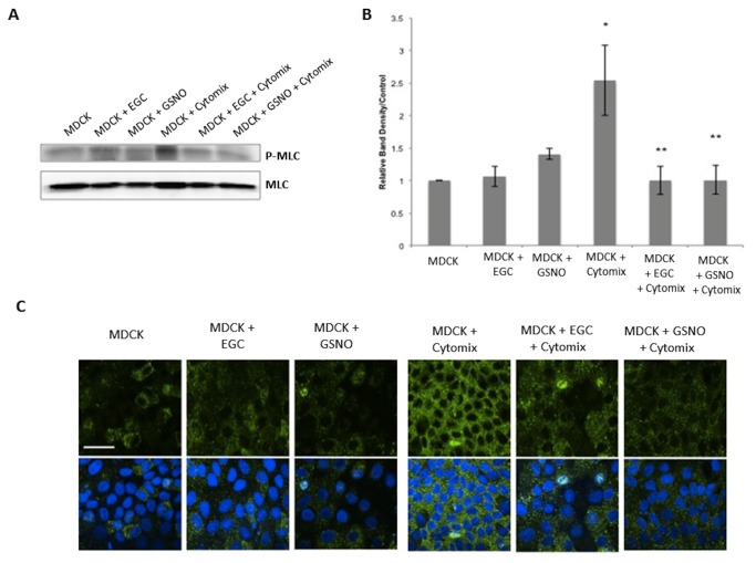
Methods: Epithelial cells were co-cultured with EGCs or incubated with GSNO and exposed to Cytomix (TNF-α, INF-γ, IL-1β) for 24 hours. Barrier function was assessed by permeability to 4kDa FITC-Dextran. Changes in tight junction proteins ZO-1, occludin, and phospho-MLC (P-MLC) were assessed by immunohistochemistry and immunoblot.

Key results: Co-culture of Cytomix-stimulated epithelial monolayers with EGCs prevented increases in permeability and improved expression and localization of occludin, ZO-1, and P-MLC. Further, treatment of epithelial monolayers with GSNO also prevented Cytomix-induced increases in permeability and exhibited a similar improvement in expression and localization of occludin, ZO-1, and P-MLC.

Conclusions & inferences: The addition of EGCs, or their secreted mediator GSNO, prevents epithelial barrier failure after injury and improved expression of tight junction proteins. Thus, therapies that increase EGC activation, such as VNS, may be a novel strategy to limit barrier failure in patients following severe injury.


Conflict of interest statement

Competing Interests: The authors have declared that no competing interests exist.

Figures

Figure 1
Figure 1. EGCs and GSNO attenuate Cytomix-induced monolayer permeability in Caco-2 cells.
Caco-2 cells were grown in either the presence or absence of EGCs or GSNO and incubated with Cytomix (TNF-α, IFN-γ, IL-1β) or PBS for 24 hours. L-NAME was used to block GSNO activity from EGCs. Caco-2 monolayer permeability to 4kDa FITC-Dextran was measured (n ≥ 4 samples per group). Cytomix-stimulation results in an increase in monolayer permeability, indicating barrier dysfunction. The presence of either EGCs or GSNO significantly reduces permeability levels. *p < 0.05 versus the controls Alone, + EGC, + GSNO ; **p < 0.01 versus + Cytomix, and +EGC +L-NAME +Cytomix.
Figure 2
Figure 2. EGCs and GSNO improve localization of ZO-1 in Caco-2 and MDCK cells.
Epithelial cells were grown in either the presence or absence of EGCs or GSNO and incubated with Cytomix (TNF-α, IFN-γ, IL-1β) for PBS for 24 hours. A: Caco-2 monolayers stained with anti-ZO-1 antibodies (green) and DAPI (blue) and imaged through confocal microscopy. Cytomix-stimulated monolayers have an altered localization of ZO-1 away from the cell surface compared to controls, indicating tight junction disruption. Stimulated cells co-cultured with EGCs or incubated with GSNO exhibited a restoration of ZO-1 localization at the cell surface. B: MDCK cells stained with anti-ZO-1 antibodies (green) and DAPI (blue) and imaged through confocal microscopy. Cytomix-stimulated monolayers have an altered localization of ZO-1 indicating tight junction disruption. Stimulated cells co-cultured with EGCs or incubated with GSNO demonstrated normal distribution of ZO-1. Images are of 60x magnification and exposure matched. Bar = 30µm.
Figure 3
Figure 3. EGCs and GSNO improve expression and localization of occludin in Caco-2 cells.
AB: Caco-2 occludin immunoblot and relative band densities. Cytomix-stimulation decreased occludin expression compared to controls. Co-culture of stimulated-cells with EGCs or incubation with GSNO prevented the Cytomix-induced loss of occludin expression. C: Caco-2 monolayers stained with anti-occludin antibodies (green) and DAPI (blue) and imaged through confocal microscopy. Cytomix-stimulated monolayers have an altered localization of occludin away from the cell surface compared to controls, indicating tight junction disruption. Stimulated cells co-cultured with EGCs or incubated with GSNO exhibited a restoration of occludin localization at the cell surface. Images are of 60x magnification and exposure matched. Bar = 30µm. *p < 0.05 versus Caco-2 cells alone, Caco-2 + EGC, Caco-2 + GSNO; **p < 0.05 versus Caco-2 + Cytomix using analysis of variance (ANOVA).
Figure 4
Figure 4. EGCs and GSNO improve expression and localization of occludin in MDCK cells.
AB: MDCK occludin immunoblot and relative band densities. Cytomix-stimulation decreased occludin expression compared to controls. Co-culture of stimulated-cells with EGCs or incubation with GSNO prevented the Cytomix-induced loss of occludin expression. C: Caco-2 monolayers stained with anti-occludin antibodies (green) and DAPI (blue) and imaged through confocal microscopy. Cytomix-stimulated monolayers have an altered localization of occludin away from the cell surface compared to controls, indicating tight junction disruption. Stimulated cells co-cultured with EGCs or incubated with GSNO exhibited a restoration of occludin localization at the cell surface. Images are of 60x magnification and exposure matched. Bar = 30µm. *p < 0.05 versus MDCK cells alone, MDCK + EGC, MDCK + GSNO ; **p < 0.05 versus MDCK + Cytomix using analysis of variance (ANOVA).
Figure 5
Figure 5. EGCs and GSNO reduce expression of P-MLC in Caco-2 cells.
AB: Caco-2 P-MLC immunoblot and relative band densities. Cytomix-stimulation increased P-MLC expression compared to controls. Co-culture of stimulated-cells with EGCs or incubation with GSNO prevented the Cytomix-induced increase in P-MLC expression. C: Caco-2 monolayers stained with anti-P-MLC antibodies (green) and DAPI (blue) and imaged through confocal microscopy. Cytomix-stimulated monolayers have increased levels of P-MLC compared to controls, indicating tight junction disruption. Co-culture of Cytomix-stimulated cells with EGCs or incubation with GSNO restores normal levels of P-MLC. Images are of 60x magnification and exposure matched. Bar = 30µm. *p < 0.05 versus Caco-2 cells alone, Caco-2 + EGC, Caco-2 + GSNO; **p < 0.05 versus Caco-2 + Cytomix using analysis of variance (ANOVA).
Figure 6
Figure 6. EGCs and GSNO reduce expression of P-MLC in MDCK cells.
AB: MDCK P-MLC immunoblot and relative band densities. Cytomix-stimulation increased P-MLC expression compared to controls. Co-culture of stimulated-cells with EGCs or incubation with GSNO prevented the Cytomix-induced increase in P-MLC expression. C: MDCK monolayers stained with anti-P-MLC antibodies (green) and DAPI (blue) and imaged through confocal microscopy. Cytomix-stimulated monolayers have increased levels of P-MLC compared to controls, indicating tight junction disruption. Co-culture of Cytomix-stimulated cells with EGCs or incubation with GSNO restores normal levels of P-MLC. Images are of 60x magnification and exposure matched. Bar = 30µm. *p < 0.05 versus MDCK cells alone, MDCK + EGC, MDCK + GSNO; **p < 0.05 versus MDCK + Cytomix using analysis of variance (ANOVA).

Similar articles

Cited by

References

    1. Musch MW, Walsh-Reitz MM, Chang EB (2006) Roles of ZO-1, occludin, and actin in oxidant-induced barrier disruption. Am J Physiol Gastrointest Liver Physiol 290: G222-G231. doi:10.1152/ajpgi.00301.2005. PubMed: 16239402. - DOI - PubMed
    1. Madara JL, Moore R, Carlson S (1987) Alteration of intestinal tight junction structure and permeability by cytoskeletal contraction. Am J Physiol 253: C854-C861. PubMed: 3425707. - PubMed
    1. Shen L, Black ED, Witkowski ED, Lencer WI, Guerriero V et al. (2006) Myosin light chain phosphorylation regulates barrier function by remodeling tight junction structure. J Cell Sci 119: 2095-2106. doi:10.1242/jcs.02915. PubMed: 16638813. - DOI - PubMed
    1. Deitch EA, Shi HP, Lu Q, Feketeova E, Skurnick J et al. (2004) Mesenteric lymph from burned rats induces endothelial cell injury and activates neutrophils. Crit Care Med 32: 533-538. doi:10.1097/01.CCM.0000109773.00644.F4. PubMed: 14758175. - DOI - PubMed
    1. Krzyzaniak MJ, Peterson CY, Cheadle G, Loomis W, Wolf P et al. (2011) Efferent vagal nerve stimulation attenuates acute lung injury following burn: The importance of the gut-lung axis. Surgery 150: 379-389. doi:10.1016/j.surg.2011.06.008. PubMed: 21783215. - DOI - PMC - PubMed

Publication types