

Neural pathways conveying novisual information to the visual cortex
- PMID: 23840972
- PMCID: PMC3690246
- DOI: 10.1155/2013/864920
Neural pathways conveying novisual information to the visual cortex
Abstract
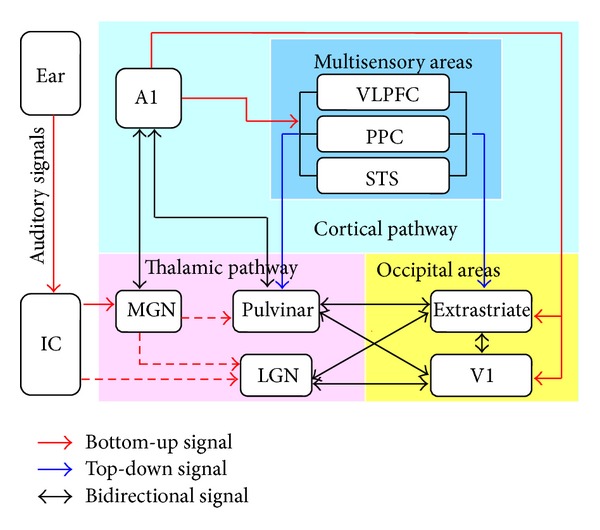
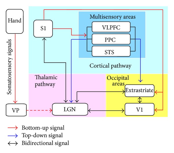
The visual cortex has been traditionally considered as a stimulus-driven, unimodal system with a hierarchical organization. However, recent animal and human studies have shown that the visual cortex responds to non-visual stimuli, especially in individuals with visual deprivation congenitally, indicating the supramodal nature of the functional representation in the visual cortex. To understand the neural substrates of the cross-modal processing of the non-visual signals in the visual cortex, we firstly showed the supramodal nature of the visual cortex. We then reviewed how the nonvisual signals reach the visual cortex. Moreover, we discussed if these non-visual pathways are reshaped by early visual deprivation. Finally, the open question about the nature (stimulus-driven or top-down) of non-visual signals is also discussed.
Figures
Similar articles
-
Coupling between Theta Oscillations and Cognitive Control Network during Cross-Modal Visual and Auditory Attention: Supramodal vs Modality-Specific Mechanisms.PLoS One. 2016 Jul 8;11(7):e0158465. doi: 10.1371/journal.pone.0158465. eCollection 2016. PLoS One. 2016. PMID: 27391013 Free PMC article. Clinical Trial.
-
Distinct effects of trial-driven and task Set-related control in primary visual cortex.Neuroimage. 2015 Oct 15;120:285-297. doi: 10.1016/j.neuroimage.2015.07.005. Epub 2015 Jul 9. Neuroimage. 2015. PMID: 26163806 Free PMC article.
-
Spatial imagery relies on a sensory independent, though sensory sensitive, functional organization within the parietal cortex: a fMRI study of angle discrimination in sighted and congenitally blind individuals.Neuropsychologia. 2015 Feb;68:59-70. doi: 10.1016/j.neuropsychologia.2015.01.004. Epub 2015 Jan 6. Neuropsychologia. 2015. PMID: 25575449
-
Mechanisms of visual attention in the human cortex.Annu Rev Neurosci. 2000;23:315-41. doi: 10.1146/annurev.neuro.23.1.315. Annu Rev Neurosci. 2000. PMID: 10845067 Review.
-
Crossmodal influences on visual perception.Phys Life Rev. 2010 Sep;7(3):269-84. doi: 10.1016/j.plrev.2010.04.006. Epub 2010 Apr 22. Phys Life Rev. 2010. PMID: 20447880 Review.
Cited by
-
Other ways of seeing: From behavior to neural mechanisms in the online "visual" control of action with sensory substitution.Restor Neurol Neurosci. 2016;34(1):29-44. doi: 10.3233/RNN-150541. Restor Neurol Neurosci. 2016. PMID: 26599473 Free PMC article. Review.
-
Using structural and functional brain imaging to uncover how the brain adapts to blindness.Ann Neurosci Psychol. 2015;2:5. Epub 2015 Aug 13. Ann Neurosci Psychol. 2015. PMID: 30288502 Free PMC article.
-
Neuroanatomical Alterations in Patients with Early Stage of Unilateral Pulsatile Tinnitus: A Voxel-Based Morphometry Study.Neural Plast. 2018 Feb 28;2018:4756471. doi: 10.1155/2018/4756471. eCollection 2018. Neural Plast. 2018. PMID: 29681925 Free PMC article.
-
Does shape discrimination by the mouth activate the parietal and occipital lobes? - near-infrared spectroscopy study.PLoS One. 2014 Oct 9;9(10):e108685. doi: 10.1371/journal.pone.0108685. eCollection 2014. PLoS One. 2014. PMID: 25299397 Free PMC article.
-
Visual deprivation selectively reshapes the intrinsic functional architecture of the anterior insula subregions.Sci Rep. 2017 Mar 30;7:45675. doi: 10.1038/srep45675. Sci Rep. 2017. PMID: 28358391 Free PMC article.
References
-
- Grill-Spector K, Malach R. The human visual cortex. Annual Review of Neuroscience. 2004;27:649–677. - PubMed
-
- Wandell BA, Dumoulin SO, Brewer AA. Visual field maps in human cortex. Neuron. 2007;56(2):366–383. - PubMed
-
- Felleman DJ, Van Essen DC. Distributed hierarchical processing in the primate cerebral cortex. Cerebral Cortex. 1991;1(1):1–47. - PubMed
-
- Seymour K, Clifford CWG, Logothetis NK, Bartels A. Coding and binding of color and form in visual cortex. Cerebral Cortex. 2010;20(8):1946–1954. - PubMed
Publication types
MeSH terms
LinkOut - more resources
Full Text Sources
Other Literature Sources
