

Primary pericardial mesothelioma: a rare entity
- PMID: 23840993
- PMCID: PMC3697233
- DOI: 10.1155/2013/283601
Primary pericardial mesothelioma: a rare entity
Abstract
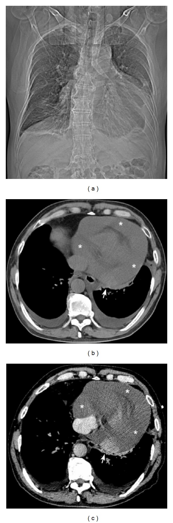
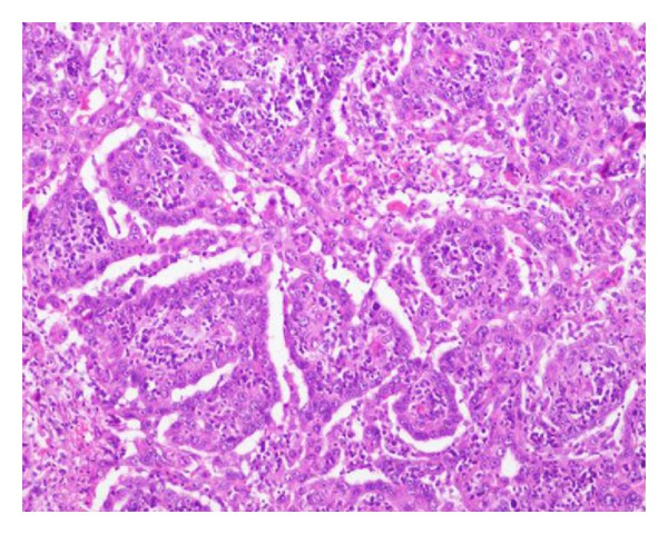
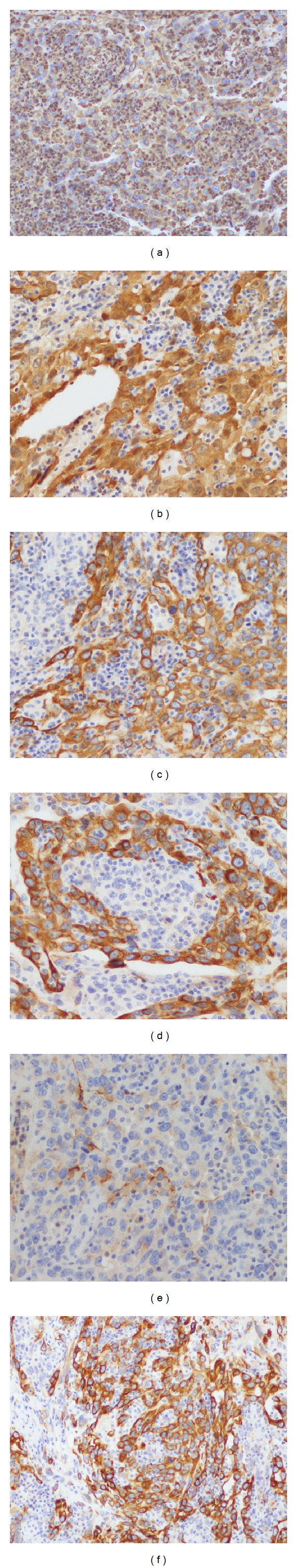
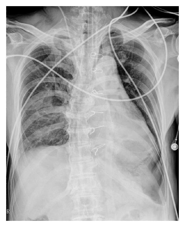
Primary pericardial malignant mesothelioma is an extremely rare neoplasm that arises from the pericardial mesothelial cell layers. Clinical symptoms and signs are frequently nonspecific, and the diagnosis is usually made after surgery or at autopsy. There is no standard treatment for pericardial mesothelioma; nonetheless, radical surgery is the mainstay of therapy for localized disease. The neoplasm is highly aggressive and carries a dismal prognosis with an overall survival of less than six months. This paper presents a case study of a 68-year-old patient with a primary pericardial malignant mesothelioma. Radiologic evaluation revealed a small nodule in the posterior pericardium with pericardial and bilateral pleural effusions. The diagnosis was established after surgery by histological and immunohistochemical studies. The patient remained alive and free of disease for about 24 months; however, due to rapid local recurrence, the patient died 27 months after the surgical treatment.
Figures
References
-
- Gossinger HD, Siostrzonek P, Zangeneh M, et al. Magnetic resonance imaging findings in a patient with pericardial mesothelioma. American Heart Journal. 1988;115(6):1321–1322. - PubMed
-
- Papi M, Genestreti G, Tassinari D, et al. Malignant pericardial mesothelioma. Report of two cases, review of the literature and differential diagnosis. Tumori. 2005;91(3):276–279. - PubMed
-
- Luk A, Ahn E, Vaideeswar P, Butany JW. Pericardial tumors. Seminars in Diagnostic Pathology. 2008;25(1):47–53. - PubMed
LinkOut - more resources
Full Text Sources
Other Literature Sources
