

doi: 10.1155/2013/456863.
Epub 2013 Jun 9.
Abdominal wall schwannoma: case report and review of the literature
Affiliations
- PMID: 23841008
- PMCID: PMC3690636
- DOI: 10.1155/2013/456863
Item in Clipboard
Abdominal wall schwannoma: case report and review of the literature
Case Rep Radiol.
2013.
Abstract
A 29-year-old female had presented to surgical outpatient's department complaining of lump in the anterior abdominal wall. Ultrasound and magnetic resonance imaging revealed a solid degenerated tumor in the anterior abdominal wall. It was surgically excised, and histopathology confirmed it to be "ancient" schwannoma. To our knowledge, this is the second reported case of an abdominal wall ancient schwannoma in the medical literature.
Figures

(a) Color flow showing hypovascularity of the mass. (b) B-mode ultrasound shows the well-encapsulated mass in anterior abdominal wall (arrow).
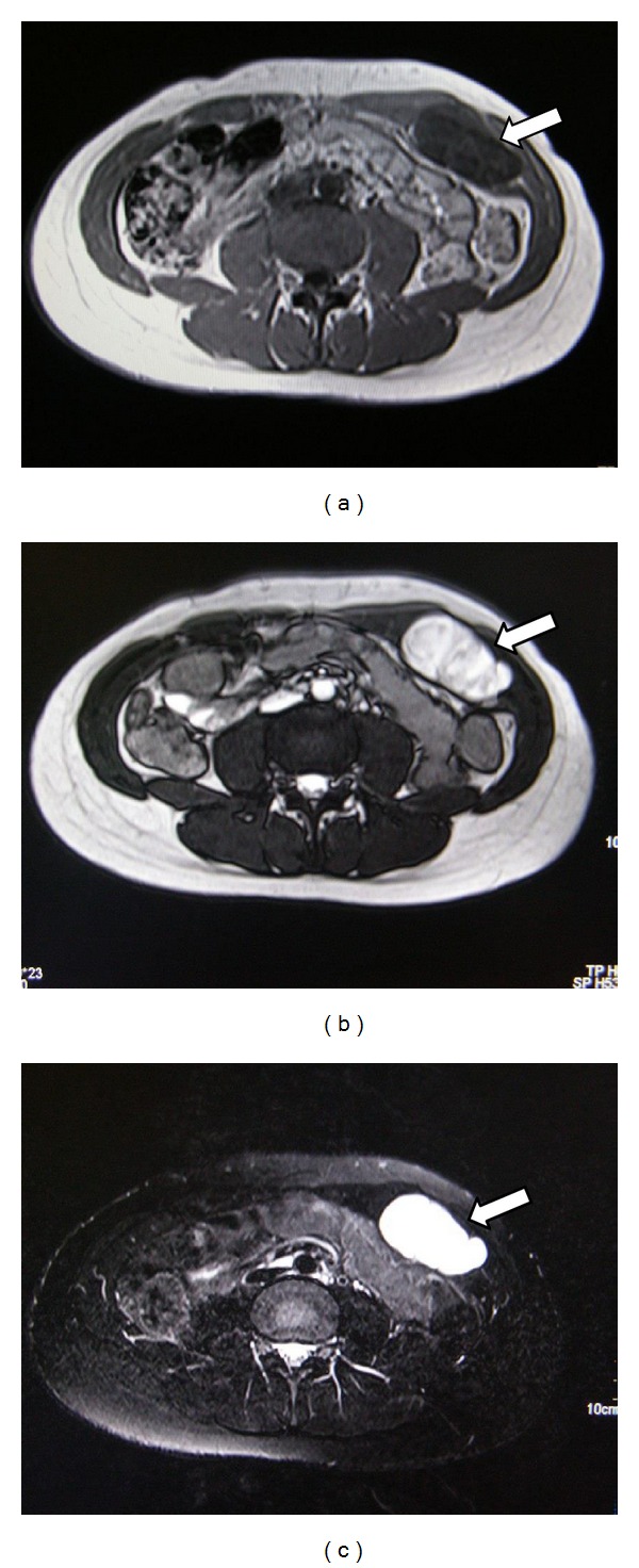
(a) T1W MRI shows encapsulated superficial mass with marked heterogeneity and patchy hypointense foci (arrow) suggesting cystic degeneration. (b) T2W MRI clearly delineates the capsule of the lesion and internal morphology (arrow). (c) The lesion is markedly hyperintense on fat-suppressed MRI suggesting degeneration (arrow).

(a) Peroperative picture shows the superficial location of the mass (arrow). (b) Excised tumor.
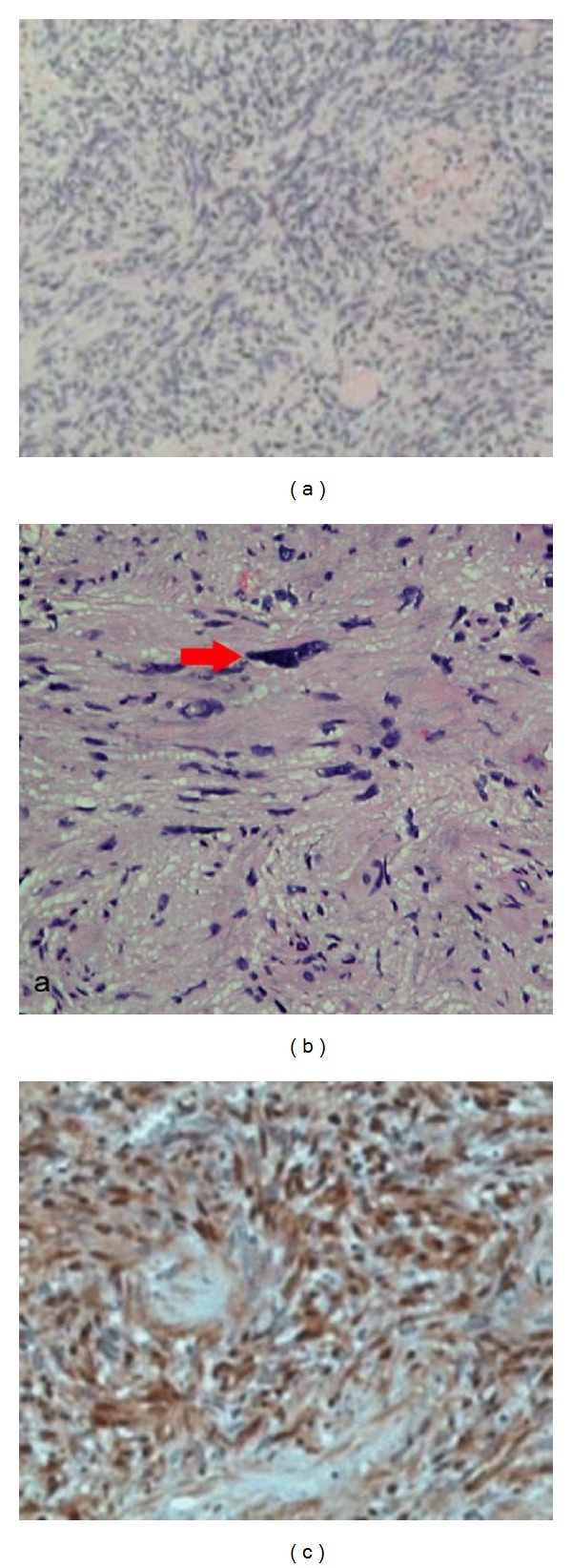
(a) Microscopic appearance of tumour showing Antoni A pattern with nuclear palisading (H & E staining). (b) Spindle cells in an Antoni type A area; red arrow indicates a large bizarre hyperchromatic nucleus. (c) Immunohistology slide confirming the diagnosis of schwannoma.
References
-
- White W, Shiu MH, Rosenblum MK, Erlandson RA, Woodruff JM. Cellular schwannoma. A clinicopathologic study of 57 patients and 58 tumors. Cancer. 1990;66(6):1266–1275. - PubMed
-
- Park MK, Lee KT, Choi YS, et al. A case of benign schwannoma in the porta hepatis. The Korean Journal of Gastroenterology. 2006;47(2):164–167. - PubMed
-
- Dane B, Dane C, Basaran S, Erginbas M, Cetin A. Vaginal Schwannoma in a case with uterine myoma. Annals of Diagnostic Pathology. 2010;14(2):137–139. - PubMed
LinkOut - more resources
Full Text Sources
Other Literature Sources
