Unicentric Castleman's Disease Arising from an Intrapulmonary Lymph Node
- PMID: 23841009
- PMCID: PMC3691905
- DOI: 10.1155/2013/289089
Unicentric Castleman's Disease Arising from an Intrapulmonary Lymph Node
Abstract
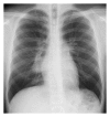
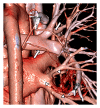
Castleman's disease is an uncommon lymphoproliferative disorder of unknown etiology, most often involving the mediastinum. It has 2 distinct clinical forms: unicentric and multicentric. Unicentric Castleman's disease arising from an intrapulmonary lymph node is rare, and establishing a preoperative diagnosis of this disease is very difficult mainly due to a lack of specific imaging features. We report a case of intrapulmonary unicentric Castleman's disease in an asymptomatic 19-year-old male patient who was accurately diagnosed by preoperative computed tomography (CT). The mass was incidentally found on a routine chest X-ray. A subsequent dynamic CT showed a well-defined, hypervascular, soft-tissue mass with small calcifications located in the perihilar area of the right lower lung. Three-dimensional CT (3D-CT) angiography indicated that the mass was receiving its blood supply through a vascular network at its surface that originated from 2 right bronchial arteries. The clinical history and CT findings were consistent with a diagnosis of unicentric Castleman's disease, and we safely and successfully removed the tumor via video-assisted thoracoscopic surgical lobectomy. This case shows that the imaging characteristics of these rare tumors on contrast-enhanced CT combined with 3D-CT angiography can be helpful in reliably establishing a correct preoperative diagnosis.
Figures


Similar articles
-
Successful Treatment of Mediastinal Unicentric Castleman's Disease Using Video-Assisted Thoracoscopic Surgery with Preoperative Embolization.Case Rep Med. 2013;2013:354507. doi: 10.1155/2013/354507. Epub 2013 Sep 30. Case Rep Med. 2013. PMID: 24198836 Free PMC article.
-
Hyaline vascular-type Castleman's disease in the hepatoduodenal ligament: report of a case.Surg Today. 2006;36(7):647-50. doi: 10.1007/s00595-006-3200-2. Surg Today. 2006. PMID: 16794803
-
Isolated intrapulmonary Castleman's disease: a case report, review of the literature.Ann Thorac Cardiovasc Surg. 2014;20 Suppl:689-91. doi: 10.5761/atcs.cr.13-00253. Epub 2014 Apr 18. Ann Thorac Cardiovasc Surg. 2014. PMID: 24747548 Review.
-
[Unusual gluteal localization of unicentric Castleman's disease: A case report and review of the literature].Ann Pathol. 2024 Mar;44(2):130-136. doi: 10.1016/j.annpat.2023.09.005. Epub 2023 Oct 3. Ann Pathol. 2024. PMID: 37798152 Review. French.
-
Rare presentation of unicentric Castleman's disease in the parotid gland.J Oral Maxillofac Surg. 2012 Sep;70(9):2114-7. doi: 10.1016/j.joms.2011.09.042. Epub 2012 Feb 2. J Oral Maxillofac Surg. 2012. PMID: 22305675
Cited by
-
Tumor enucleation for Castleman's disease in the pulmonary hilum: a case report.Surg Case Rep. 2019 Jun 10;5(1):95. doi: 10.1186/s40792-019-0652-3. Surg Case Rep. 2019. PMID: 31183765 Free PMC article.
-
Amyloidosis secondary to intrapulmonary Castleman disease mimicking pulmonary hyalinizing granuloma-like clinical features: A rare case report.Medicine (Baltimore). 2019 Apr;98(14):e15039. doi: 10.1097/MD.0000000000015039. Medicine (Baltimore). 2019. PMID: 30946344 Free PMC article.
-
Rare Unicentric Intrapulmonary Castleman Disease: A Systematic Review and Report of a Case.Open Respir Med J. 2025 Feb 18;19:e18743064348696. doi: 10.2174/0118743064348696250107092627. eCollection 2025. Open Respir Med J. 2025. PMID: 40322498 Free PMC article.
-
Unicentric Castleman's disease presenting as a pulmonary mass: a diagnostic dilemma.Am J Case Rep. 2015 Apr 30;16:259-61. doi: 10.12659/AJCR.893380. Am J Case Rep. 2015. PMID: 25928278 Free PMC article.
-
Video-assisted thoracoscopic surgery is a safe and effective method to treat intrathoracic unicentric Castleman's disease.BMC Surg. 2020 Jun 10;20(1):127. doi: 10.1186/s12893-020-00789-6. BMC Surg. 2020. PMID: 32522182 Free PMC article.
References
-
- Castleman B, Towne VW. Case records of the Massachusetts general hospital: case 32-1984. The New England Journal of Medicine. 1954;311:388–398. - PubMed
-
- Olak J. Benign lymph node disease involving the mediastinum. In: Shields TW, LoCicero J, Reed CE, Feins RH, editors. General Thoracic Surgery. 7th edition. Vol. 2. Philadelphia, Pa, USA: Lippincott Williams & Wilkins; 2009. pp. 2366–2367.
-
- Herrada J, Cabanillas FF. Multicentric Castleman’s disease. The American Journal of Clinical Oncology. 1995;18(2):180–183. - PubMed
-
- Keller AR, Hochholzer L, Castleman B. Hyaline-vascular and plasma-cell types of giant lymph node hyperplasia of the mediastinum and other locations. Cancer. 1972;29(3):670–683. - PubMed
-
- Mohanna S, Sanchez J, Ferrufino JC, Bravo F, Gotuzzo E. Characteristics of Castleman’s disease in Peru. European Journal of Internal Medicine. 2006;17(3):170–174. - PubMed
LinkOut - more resources
Full Text Sources
Other Literature Sources

