

Intraosseous Ganglia: a series of 17 treated cases
- PMID: 23841071
- PMCID: PMC3697270
- DOI: 10.1155/2013/462730
Intraosseous Ganglia: a series of 17 treated cases
Abstract
Background: Intraosseous ganglion is a cystic lesion that contains gelatinous material, most often occurs in middle-aged patients, and is regarded as similar to soft-tissue ganglion. The etiology is unknown, but association with degenerative joint disease has been considered.
Materials and methods: At a single institute, 17 patients (8 men, 9 women) with a mean age of 48.9 years (22-72 years) were surgically treated for an intraosseous ganglion. The lesions were located in 9 long bones (5 tibiae, 2 humeri, 1 ulna, and 1 femur); 4 flat bones (2 scapulae, 2 ilia); and 4 small bones (2 scaphoid, 1 metacarpal bone, and 1 talus). The diagnosis was confirmed based both on the gross intraoperative finding of intralesional gelatinous material and on histopathology.
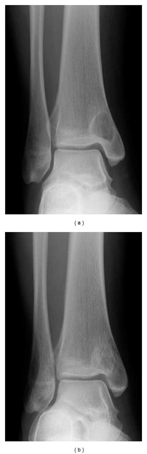
Results: All lesions occurred at the epiphysis or near the joint. The plain radiographs showed a lesion with marginal osteosclerosis. The average lesion size was 22.4 mm (range 6-40 mm). Among the 17 patients, 2 (12%) had osteoarthritis, 3 (18%) had pathological fracture, and 4 (24%) had extraskeletal extension.
Discussion and conclusion: The periosteum and cortex of bone represent physical barriers. Therefore, it seems much more likely that primary bone lesions will spread to the soft tissues. Intraosseous ganglion does not appear to be associated with either soft-tissue ganglion or with osteoarthritis. This clinical information and the appearance on plain radiographs, particularly the marginal osteosclerosis, are of differential diagnostic importance.
Figures
References
-
- FISK GR. Bone concavity caused by a ganglion. The Journal of Bone and Joint Surgery. 1949;31(2):220–221. - PubMed
-
- Crabbe WA. Intra-osseous ganglia of bone. British Journal of Surgery. 1966;53(1):15–17. - PubMed
-
- Feldman F, Johnston A. Intraosseous ganglion. American Journal of Roentgenology, Radium Therapy, and Nuclear Medicine. 1973;118(2):328–343. - PubMed
-
- Dorfman HD, Czerniak B. Bone Tumors. Mosby Year Book; 1998.
-
- Schajowicz F, Clavel Sainz M, Slullitel JA. Juxta-articular bone cysts (intra-osseous ganglia). A clinicopathological study of eighty-eight cases. Journal of Bone and Joint Surgery B. 1979;61(1):107–116. - PubMed
Publication types
MeSH terms
LinkOut - more resources
Full Text Sources
Other Literature Sources
Medical
