Review
. 2013 Jul;8(7):855-75.
doi: 10.2217/fmb.13.52.

Progress in targeting cell envelope biogenesis in Mycobacterium tuberculosis

Affiliations
Review

Progress in targeting cell envelope biogenesis in Mycobacterium tuberculosis

Mary Jackson et al. Future Microbiol. 2013 Jul.

Abstract

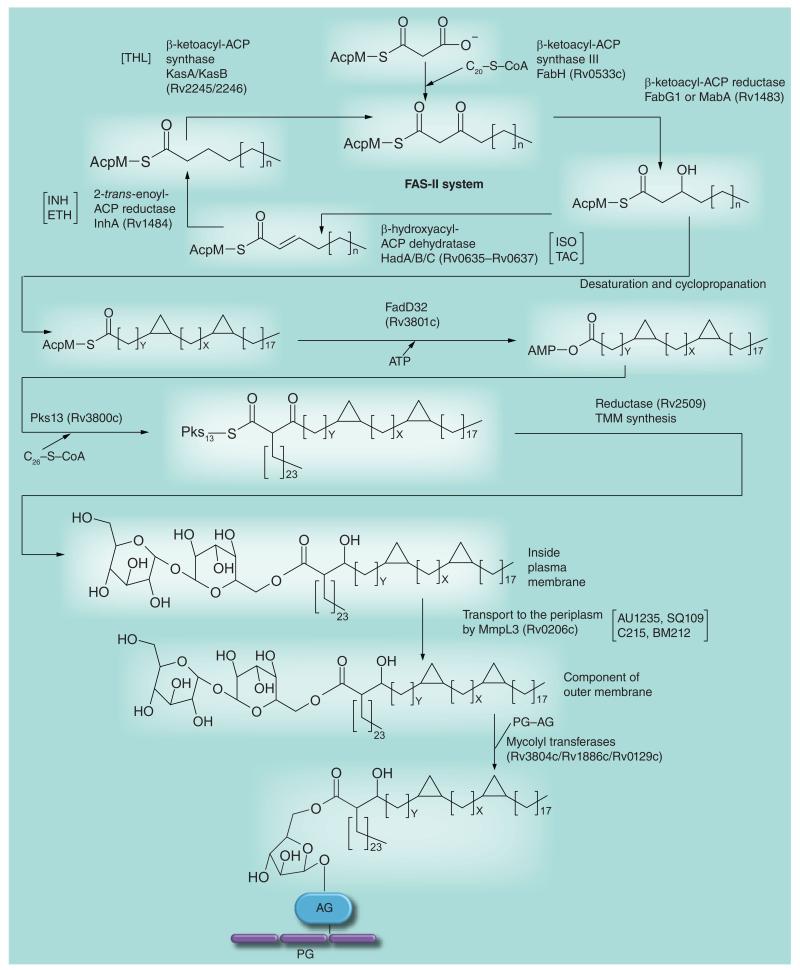
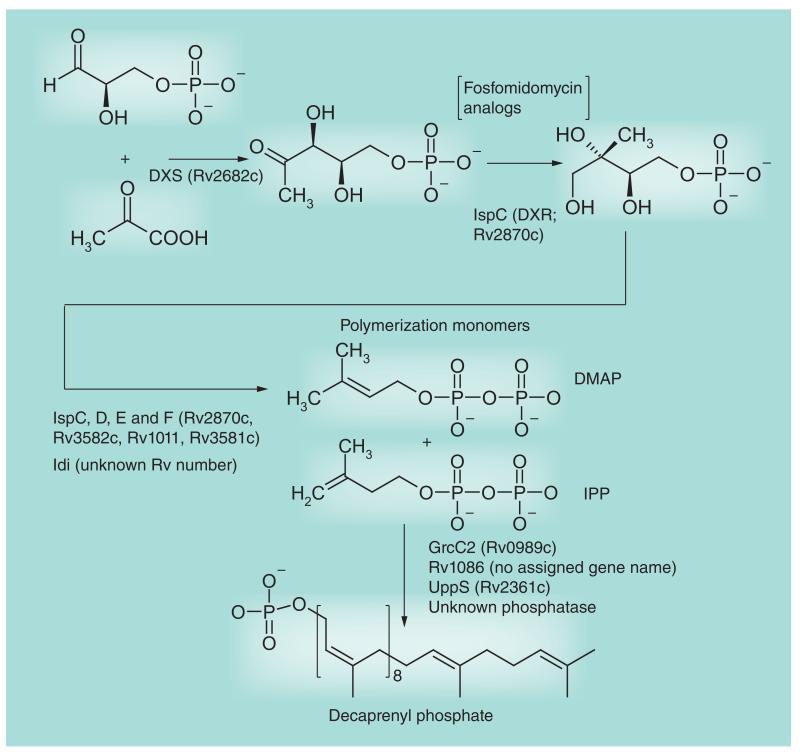
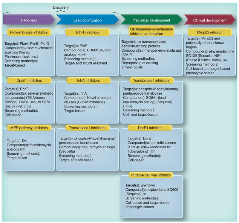
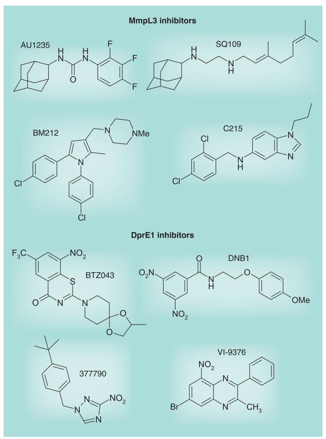
Most of the newly discovered compounds showing promise for the treatment of TB, notably multidrug-resistant TB, inhibit aspects of Mycobacterium tuberculosis cell envelope metabolism. This review reflects on the evolution of the knowledge that many of the front-line and emerging products inhibit aspects of cell envelope metabolism and in the process are bactericidal not only against actively replicating M. tuberculosis, but contrary to earlier impressions, are effective against latent forms of the disease. While mycolic acid and arabinogalactan synthesis are still primary targets of existing and new drugs, peptidoglycan synthesis, transport mechanisms and the synthesis of the decaprenyl-phosphate carrier lipid all show considerable promise as targets for new products, older drugs and new combinations. The advantages of whole cell- versus target-based screening in the perpetual search for new targets and products to counter multidrug-resistant TB are discussed.


Figures

Figure 1
Figure 1. A depiction of the Mycobacterium tuberculosis cell envelope
The cell wall core (also known as mycolyl AG–PG or mAGP) consists of the PG (black with colored peptides extending from it) attached to the mycolate layer (inner leaflet of the outer membrane) via the AG. The galactofuranosyl residues are shown in blue and the arabinofuranosyl residues in red. The mycolic acids attached to the arabinan are shown in black and are in a folded ‘W’ configuration; the pink mycolic acids are attached to trehalose (trehalose monomycolate with a single mycolic acid and trehalose dimycolate with two mycolic acids) and are expected to be found in both leaflets of the outer membrane. Lipoarabinomannan (arabinofuranosyl residues in red as in AG and mannopyranosyl residues in purple) is shown both in the outer membrane and plasma membrane as is lipomannan. Phospholipids are only shown in the plasma membrane, but are also found in the outer membrane. The capsule is shown containing glucan (black), mannan (purple) and arabinomannan (purple and red). The dimensions of the various layers are from published electron micrographic studies [112-114]. Molecular modeling suggests that the length of the medial zone could be as large as 20 nm; the arabinan and galactan are quite flexible and a very dense contracted configuration is required for the much smaller 6.5-nm medial zone suggested by the electron microscopic studies; this perhaps could change under different growth conditions. The cell wall core (left side of figure) is the primary target discussed in this review. Please see full size image as supplementary Figure 1 online at www.futuremedicine.com/doi/suppl/10.2217/fmb.13.52. AG: Arabinogalactan; PG: Peptidoglycan.
Figure 2
Figure 2. Biosynthesis of cell wall rhamnosyl residues and the formation of lipid-linked galactan
This figure focuses on the targeted enzymes, RmlC, RmlD and Glf, but includes all of the enzymes involved in making the linker and galactan regions of arabinogalactan. As with the early steps of peptidoglycan biosynthesis, a large number of potential drug target enzymes (eight in total) are present. RmlA–D are soluble enzymes involved in making dTDP-Rha; Glf is a soluble enzyme that makes UDP-Galf. WbbL, GlfT1 and GlfT2 are glycosyltransferases that use the dTDP-Rha and UDP-Galf, respectively, to attach Rha and Galf to the lipid intermediate shown. WecA is a GlcNAc-phosphate transferase that attaches GlcNAc-1-P to decaprenyl phosphate. For further details of the pathway, see [33].
Figure 3
Figure 3. Arabinosyl biosynthesis
This figure focuses on the targeted enzymes DprE1 and EmbA, B and C. The activated donor of arabinosyl residues, decaprenyl phosphoryl arabinose, is formed from the nucleotide biosynthetic intermediate phosphoribose pyrophosphate in four enzymatic steps beginning with the transfer of phosphoribose to decaprenyl phosphate. Decaprenyl phosphoryl arabinose is then used as the arabinosyl donor by the arabinosyltransferases. The final product of this figure, AG attached to PG, is then mycolylated (see Figure 5) to form the complete cell wall core (mAGP). The sites of action of the inhibitors mentioned in this review are indicated between square brackets. AG: Arabinogalactan; EMB: Ethambutol; PG: Peptidoglycan.
Figure 4
Figure 4. Peptidoglycan biosynthesis
This figure of the pathway focuses on enzyme targets discussed in this review. The pathway divides into the cytoplasmic formation of Park’s nucleotide (see top and middle of figure) and then the formation of the membrane-bound decaprenyl diphosphate Mur pentapeptide followed by polymerization, cross-linking and trimming. The early enzymatic steps forming Park’s nucleotide appear to be fertile for drug targeting, but as discussed in the text, they have only yielded d-cycloserine, although new work continues. The phospho-N-acetylmuramyl pentapeptide translocase MraY is thought to be the target of capuramycin analogs currently undergoing preclinical development. The cross-linking event late in the pathway is the target for the β-lactams that are now receiving attention for use against Mycobacterium tuberculosis. For further details on the formation of peptidoglycan, see [33]. DAP: Diaminopimelic acid; Glyc: Glycolate; PG: Peptidoglycan.
Figure 5
Figure 5. Mycolic acid biosynthesis
This figure of the pathway focuses on targets discussed in this review. The meromycolate carbon chain is formed via the FAS-II system (top of figure). The introduction of the double bonds is not yet understood; it may involve an isomerization event during FAS-II elongation or occur (as shown in the figure) after formation of the carbon chain. After cyclopropanation and (not shown in the figure) formation of keto and methoxy groups, the meromycolate is activated and condenses with C26-S-CoA; the C26-S-CoA is formed from the FAS-I fatty acid elongation system. After reduction, the now mature mycolic acid is attached to trehalose to form TMM by an unknown enzyme and mechanism. It is then transported to the cell envelope outside the plasma membrane and attached to AG by a transport mechanism involving MmpL3 and the mycolyl transferases, respectively. For further details of the pathway, see [33]. The sites of action of the inhibitors mentioned in this review are indicated between square brackets. AG: Arabinogalactan; ETH: Ethionamide; INH: Isoniazid; ISO: Isoxyl; PG: Peptidoglycan; TAC: Thiacetazone; THL: Thiolactomycin; TMM: Trehalose monomycolate.
Figure 6
Figure 6. Decaprenyl phosphate biosynthesis
This figure focuses on the targeted enzyme DXR (IspC). The first six enzymes, including DXR, are involved in the formation of polymerization precursors DMAP and IPP via the non-mevalonate pathway. The later enzymes catalyze the polymerization and dephosphorylation reactions. Decaprenyl phosphate is used for arabinan, arabinogalactan and peptidoglycan biosynthesis. DMAP: Dimethylallyl pyrophosphate; DXS: 1-deoxyxylulose-5-phosphate; IPP: Isopentyl pyrophosphate.
Figure 7
Figure 7. Global TB drug pipeline showing the cell envelope-related inhibitors under development
See text and the Working Group for New Drugs, Stop TB Partnership [203] for details.
Figure 8
Figure 8. Structures of MmpL3 and DprE1 inhibitors

Similar articles

Cited by

References

    1. Center for Disease Control and Prevention: emergence of Mycobacterium tuberculosis with extensive resistance to second-line drugs worldwide, 2000-2004. MMWR Morb. Mortal. Wkly Rep. 2006;55(11):301–305. - PubMed
    1. Mitchison D, Davies G. The chemotherapy of tuberculosis: past, present and future. Int. J. Tuberc. Lung Dis. 2012;16(6):724–732. - PMC - PubMed
    1. Bhamidi S, Shi L, Chatterjee D, Belisle JT, Crick DC, McNeil MR. A bioanalytical method to determine the cell wall composition of Mycobacterium tuberculosis grown in vivo. Anal. Biochem. 2012;421(1):240–249. - PubMed
    1. Ryan GJ, Hoff DR, Driver ER, et al. Multiple M. tuberculosis phenotypes in mouse and guinea pig lung tissue revealed by a dual-staining approach. PLoS One. 2010;5(6):e11108. - PMC - PubMed
    1. Cunningham AF, Spreadbury CL. Mycobacterial stationary phase induced by low oxygen tension: cell wall thickening and localization of the 16-kilodalton α-crystallin homolog. J. Bacteriol. 1998;180(4):801–808. - PMC - PubMed

Websites

    1. WHO Tuberculosis. www.who.int/mediacentre/factsheets/fs104/en.
    1. PubChem BioAssay Inhibitors of mycobacterial glucosamine-1-phosphate acetyl transferase (GlmU) – bioassay summary. http://pubchem.ncbi.nlm.nih.gov/assay/assay.cgi?aid=1376.
    1. Working Group on New TB Drugs Stop TB Partnership. www.newtbdrugs.org.

Publication types

MeSH terms

Substances