. 2013 Aug;121(4):293-302.
doi: 10.1111/eos.12059. Epub 2013 May 30.

Ameloblasts require active RhoA to generate normal dental enamel

Affiliations

Ameloblasts require active RhoA to generate normal dental enamel

Hui Xue et al. Eur J Oral Sci. 2013 Aug.

Abstract

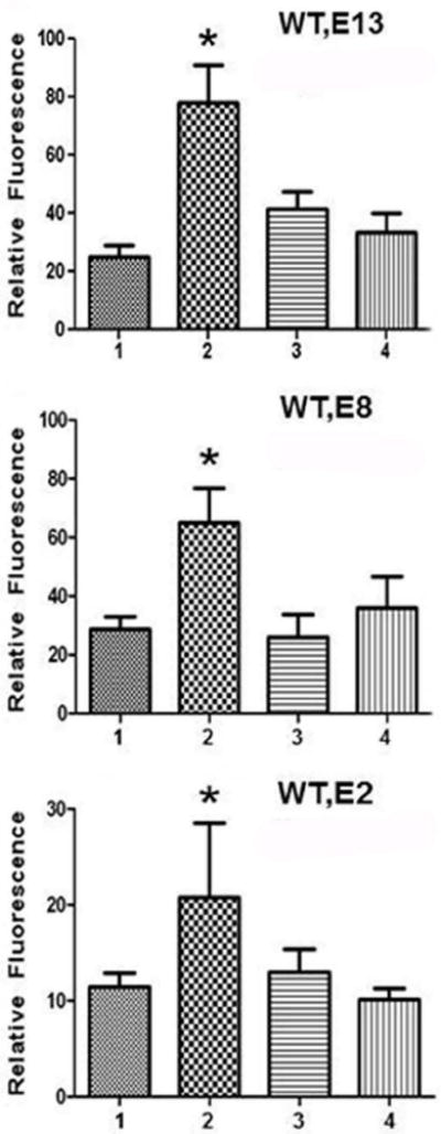
RhoA plays a fundamental role in regulation of the actin cytoskeleton, intercellular attachment, and cell proliferation. During amelogenesis, ameloblasts (which produce the enamel proteins) undergo dramatic cytoskeletal changes and the RhoA protein level is up-regulated. Transgenic mice were generated that express a dominant-negative RhoA transgene in ameloblasts using amelogenin gene-regulatory sequences. Transgenic and wild-type (WT) molar tooth germs were incubated with sodium fluoride (NaF) or sodium chloride (NaCl) in organ culture. Filamentous actin (F-actin) stained with phalloidin was elevated significantly in WT ameloblasts treated with NaF compared with WT ameloblasts treated with NaCl or with transgenic ameloblasts treated with NaF, thereby confirming a block in the RhoA/Rho-associated protein kinase (ROCK) pathway in the transgenic mice. Little difference in quantitative fluorescence (an estimation of fluorosis) was observed between WT and transgenic incisors from mice provided with drinking water containing NaF. We subsequently found reduced transgene expression in incisors compared with molars. Transgenic molar teeth had reduced amelogenin, E-cadherin, and Ki67 compared with WT molar teeth. Hypoplastic enamel in transgenic mice correlates with reduced expression of the enamel protein, amelogenin, and E-cadherin and cell proliferation are regulated by RhoA in other tissues. Together these findings reveal deficits in molar ameloblast function when RhoA activity is inhibited.

Keywords: ameloblasts; dental enamel; dominant-negative RhoA; transgenic mice.


Conflict of interest statement

CONFLICTS OF INTEREST

This work did not involve any conflicts of interest.

Figures

Fig. 1
Fig. 1. Transgene expression in developing molar teeth
Western blot of extracts of first mandibular molar teeth probed with anti-RhoA antibody. Transgenic bands are visible at approximately 48 kDa for TgEGFP-RhoADN-2, 8 and 13 (E2, E8, E13) and all mice have endogenous RhoA at 22 kDa; anti-actin was used for normalization. Quantitation from two independent experiments indicated TgEGFP-RhoADN-2 (E2) expressed 35% and TgEGFP-RhoADN-8 (E8) expressed 20% of TgEGFP-RhoADN-13 (E13) transgenic protein.
Fig. 2
Fig. 2. Immunohistochemistry localizes transgenic protein to ameloblasts during the secretory stage of enamel development
Sections from first mandibular molars from TgEGFP-RhoADN-13 mice were incubated with anti-GFP antibody at postnatal days PN1 (A), PN2 (B), PN3 (C), PN4 (D), PN6 (E) and PN8 (F). Scale bars = 100 μm.
Fig. 3
Fig. 3. Organ culture demonstrates effectiveness of the dominant negative mutation in 3 transgenic lines
First mandibular molar teeth from WT or TgEGFP-RhoADN-2, 8 or 13 (E2, E8, E13) transgenic lines were placed into organ culture and treated with 4mM NaCl or NaF for 30 min. Following phalloidin staining of sections, fluorescence relative intensities of ameloblasts were quantitated. For each comparison, bar 1: WT, NaCl; bar 2: WT, NaF; bar 3: transgenic, NaCl; bar 4: transgenic, NaF. The * indicates significant difference between WT treated with NaF and either WT treated with NaCl or transgenic treated with NaF (P<0.0001).
Fig. 4
Fig. 4. Dental fluorosis assessment of incisor teeth
Clinical images of mandibular incisors. WT (W) and TgEGFP-RhoADN-13 (E) mice were provided drinking water for 4 weeks containing 0, 50 or 100 ppm F ad libitum.
Fig. 5
Fig. 5. Quantitative fluorescence (QF) of mandibular incisors
Results are shown for each treatment/control group and genotype shown in Fig. 4. *Results significantly different from controls.
Fig. 6
Fig. 6. Western blot to compare transgene expression in mandibular molar and incisor
Extracts of PN4 transgenic teeth (10 μg per lane) were probed with anti-GFP antibody. M: molar; In: incisor.
Fig. 7
Fig. 7. Amelogenin is reduced in TgEGFP-RhoADN-13 teeth, by Western blot
A. Quantitation of normalized band intensity, where * indicates statistical difference from controls (P<0.001 at PN 2 and 4). W0 to W8 are PN ages for WT mice; E0 to E8 are PN ages for TgEGFP-RhoADN-13 mice. B. Western blot using anti-amelogenin antibody to detect amelogenin proteins from enamel organs from mice at the indicated ages; C. Normalization using anti-GAPDH for PN0 through PN8 for WT and transgenic mice.
Fig. 8
Fig. 8. E-cadherin immunostaining of ameloblasts from WT and TgEGFP-RhoADN-13 mice
Mandibular first molars (B-C) and maxillary second molars (D-E) from PN4 mice were stained with anti-E-cadherin antibody. A: First molar control lacking primary antibody; B,D: WT, anti-E-cadherin; C,E: TgEGFP-RhoADN-13, anti-E-cadherin. Scale bar = 200 μm.
Fig. 9
Fig. 9. Ki67 labels fewer ameloblast nuclei in TgEGFP-RhoADN-13 transgenic compared to WT first mandibular molars from mice of the same age
Mandibular PN3 (A,B,E) and maxillary PN2 (C,D) first molars. A,C: WT, anti-Ki67; B,D: TgEGFP-RhoADN-13, anti-Ki67; E: control lacking primary antibody. Dark nuclei represent Ki67 positive ameloblasts in magnified inserts. Scale bar = 200 μm.

References

    1. THESLEFF I, SHARPE P. Signaling networks regulating dental development. Mech Dev. 1997;67:111–123. - PubMed
    1. JERNVALL J, THESLEFF I. Reiterative signaling and patterning during mammalian tooth morphogenesis. Mech Dev. 2000;92:19–29. - PubMed
    1. SMITH CE, NANCI A. Overview of morphological changes in enamel organ cells associated with major events in amelogenesis. Int J Dev Biol. 1995;39:153–161. - PubMed
    1. SMITH CE. Ameloblasts: secretory and resorptive functions. J Dent Res. 1979;58(Spec Issue B):695–707. - PubMed
    1. SMITH CE. Cellular and chemical events during enamel maturation. Crit Rev Oral Biol Med. 1998;9:128–161. - PubMed

Publication types

MeSH terms

LinkOut - more resources