. 2013 Jul 11:11:170.
doi: 10.1186/1479-5876-11-170.

Importance of leptin signaling and signal transducer and activator of transcription-3 activation in mediating the cardiac hypertrophy associated with obesity

Affiliations

Importance of leptin signaling and signal transducer and activator of transcription-3 activation in mediating the cardiac hypertrophy associated with obesity

Maren Leifheit-Nestler et al. J Transl Med. .

Abstract

Background: The adipokine leptin and its receptor are expressed in the heart, and leptin has been shown to promote cardiomyocyte hypertrophy in vitro. Obesity is associated with hyperleptinemia and hypothalamic leptin resistance as well as an increased risk to develop cardiac hypertrophy and heart failure. However, the role of cardiac leptin signaling in mediating the cardiomyopathy associated with increased body weight is unclear, in particular, whether it develops subsequently to cardiac leptin resistance or overactivation of hypertrophic signaling pathways via elevated leptin levels.

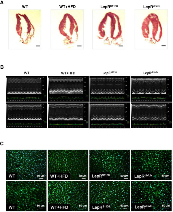
Methods: The cardiac phenotype of high-fat diet (HFD)-induced obese wildtype (WT) mice was examined and compared to age-matched genetically obese leptin receptor (LepR)-deficient (LepRdb/db) or lean WT mice. To study the role of leptin-mediated STAT3 activation during obesity-induced cardiac remodeling, mice in which tyrosine residue 1138 within LepR had been replaced with a serine (LepRS1138) were also analyzed.

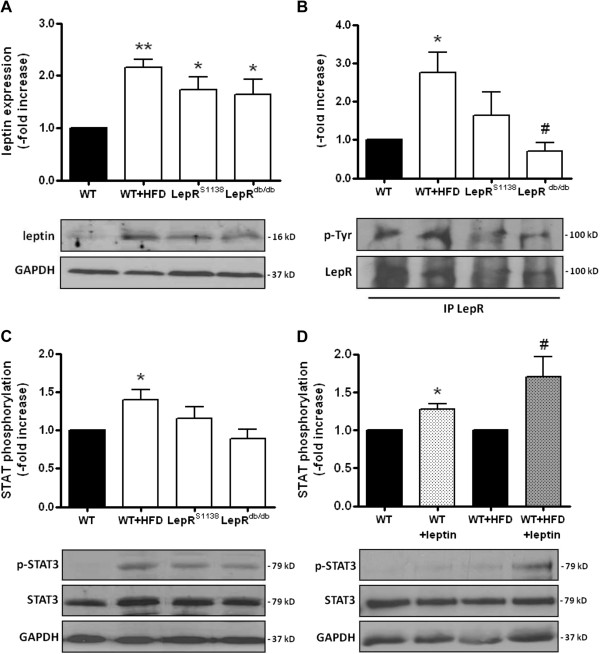
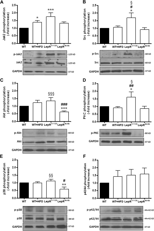
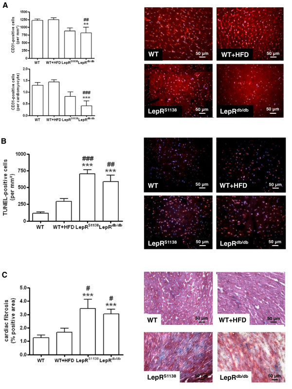
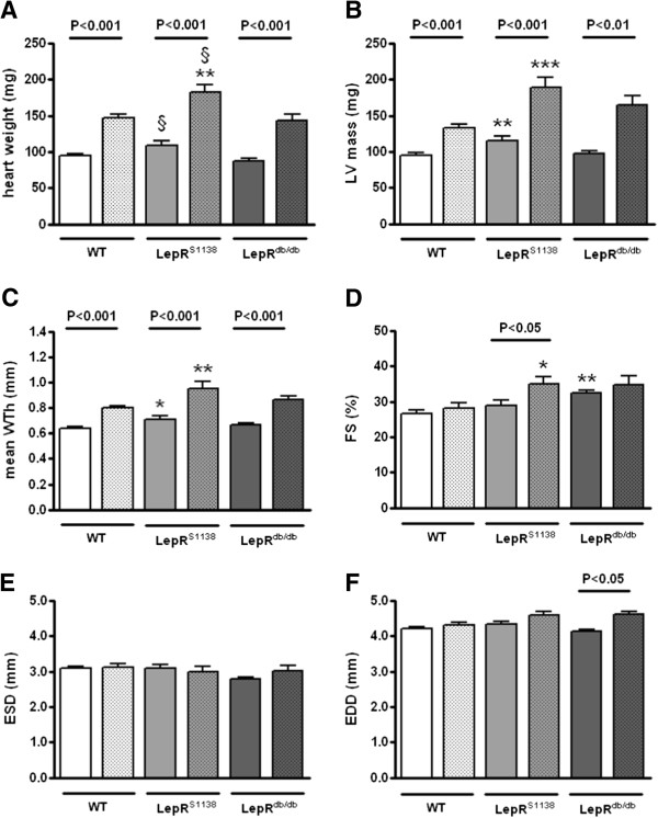
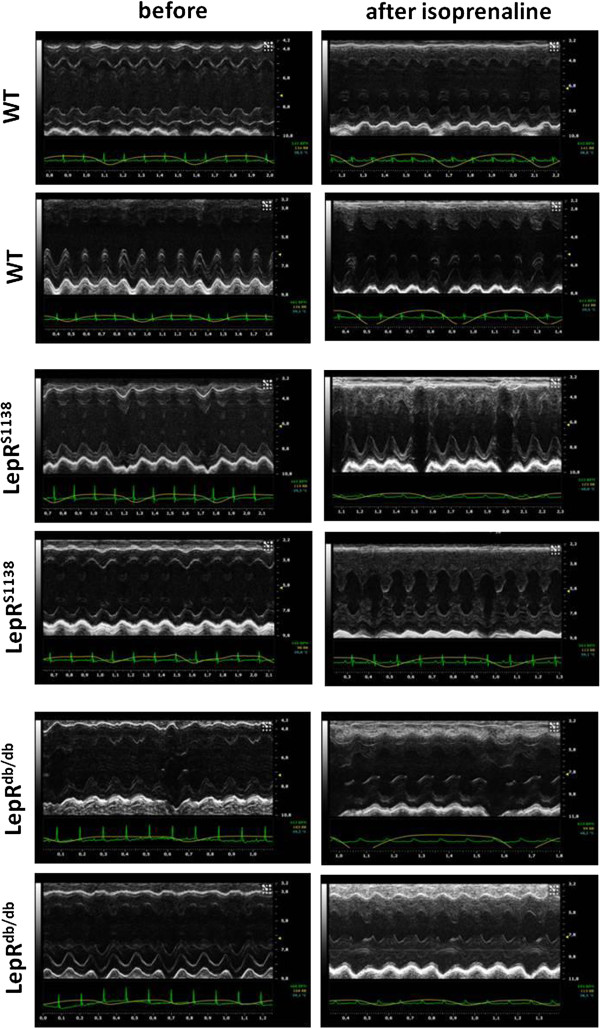
Results: Obesity was associated with hyperleptinemia and elevated cardiac leptin expression in both diet-induced and genetically obese mice. Enhanced LepR and STAT3 phosphorylation levels were detected in hearts of obese WT mice, but not in those with LepR mutations. Moreover, exogenous leptin continued to induce cardiac STAT3 activation in diet-induced obese mice. Although echocardiography revealed signs of cardiac hypertrophy in all obese mice, the increase in left ventricular (LV) mass and diameter was significantly more pronounced in LepRS1138 animals. LepRS1138 mice also exhibited an increased activation of signaling proteins downstream of LepR, including Jak2 (1.8-fold), Src kinase (1.7-fold), protein kinase B (1.3-fold) or C (1.6-fold). Histological analysis of hearts revealed that the inability of leptin to activate STAT3 in LepRdb/db and LepRS1138 mice was associated with reduced cardiac angiogenesis as well as increased apoptosis and fibrosis.

Conclusions: Our findings suggest that hearts from obese mice continue to respond to elevated circulating or cardiac leptin, which may mediate cardioprotection via LepR-induced STAT3 activation, whereas signals distinct from LepR-Tyr1138 promote cardiac hypertrophy. On the other hand, the presence of cardiac hypertrophy in obese mice with complete LepR signal disruption indicates that additional pathways also play a role.


Figures

Figure 1
Figure 1
Cardiac phenotype of lean and obese WT, WT + HFD, LepRS1138 and LepRdb/db mice. (A) Representative H&E-stained longitudinal sections through hearts of 7 months-old mice are shown. Magnification, ×10. (B) Representative M-mode echocardiographic recordings. (C) Representative images of wheat germ agglutinin (WGA)-stained myocardial cross sections. The mean cardiomyocyte cross-sectional areas are given in Table 1.
Figure 2
Figure 2
Cardiac leptin expression and signal transduction in lean and obese mice. Protein was extracted from hearts of 7 months-old mice (n = 8 per group) and analyzed for the expression of (A) leptin, (B) phosphorylated LepR (using immunoprecipitation of LepR, followed by the detection of total phosphotyrosines and LepR) and (C) phosphorylated STAT3. (D) Cardiac STAT3 phosphorylation in response (30 min later) to a single injection of recombinant murine leptin (1 mg/kg BW i.p.) was examined in WT (n = 4) and WT + HFD (n = 6) mice. Results are expressed as -fold increase of controls (black bars) after normalization for total protein and GAPDH expression. The mean ± SEM as well as representative Western blot results are shown. *P < 0.05 and **P < 0.01 vs. WT mice; #P < 0.05 vs. WT + HFD mice.
Figure 3
Figure 3
Hypertrophic signal transduction in hearts of lean and obese mice. Protein was isolated from hearts of 7 months-old WT (n = 15), WT + HFD (n = 12), LepRS1138 (n = 15) and LepRdb/db (n = 15) mice and analyzed for the expression of phosphorylated Jak2 (A), Src kinase (B), Akt (C), PKC (D), p38 (E) and p42/44 MAPK (F). Results are expressed as -fold increase of lean control mice (after normalization for total protein [with the exception of PKC] and GAPDH expression). The mean ± SEM as well as representative findings are shown. *P < 0.05, **P < 0.01 and ***P < 0.001 vs. WT mice; #P < 0.05 and ##P < 0.01 vs. WT + HFD mice; §P < 0.05, §§P < 0.01 and §§§P < 0.001 for the difference between LepRdb/db and LepRS1138 mice.
Figure 4
Figure 4
Histological analysis of angiogenesis, apoptosis and fibrosis in hearts of lean and obese mice. Serial cross sections through the LV of WT, WT + HFD, LepRS1138 and LepRdb/db mice (n = 10 per group) were immunostained and the number of (A) CD31-positive endothelial cells and (B) TUNEL-positive apoptotic cell nuclei determined. Results are expressed per cardiomyocyte and/or mm2. (C) The degree of cardiac fibrosis was quantified after Masson’s trichrome (MTC) staining. Results are expressed as % of total tissue area (at 200-fold magnification). The mean ± SEM as well as representative findings are shown. **P < 0.01 and ***P < 0.001 vs. WT; #P < 0.05, ##P < 0.01 and ###P < 0.001 vs. WT + HFD mice.
Figure 5
Figure 5
Echocardiography findings in young lean and obese mice before and after chronic β-adrenergic stimulation. Isoprenaline-filled osmotic minipumps were subcutaneously implanted into 2 months-old WT (n = 12), LepRS1138 (n = 9) and LepRdb/db (n = 7) mice to examine the cardiac response to a hypertrophic stimulus other than leptin. Echocardiography (A-F) was performed immediately before (open bars) as well as at the time of tissue harvest 14 days later (dotted bars). *P < 0.05, *P < 0.01 and ***P < 0.001 for differences vs. WT mice; §P < 0.05 for differences between LepRdb/db and LepRS1138 mice. Significance levels for differences before and after isoprenaline stimulation (as determined using Student’s t test for paired means) are indicated within the graph.
Figure 6
Figure 6
Representative M-mode echocardiography recordings.

Similar articles

Cited by

References

    1. Considine RV, Sinha MK, Heiman ML, Kriauciunas A, Stephens TW, Nyce MR, Ohannesian JP, Marco CC, McKee LJ, Bauer TL. Serum immunoreactive-leptin concentrations in normal-weight and obese humans. N Engl J Med. 1996;334:292–295. doi: 10.1056/NEJM199602013340503. - DOI - PubMed
    1. Gottdiener JS, Reda DJ, Materson BJ, Massie BM, Notargiacomo A, Hamburger RJ, Williams DW, Henderson WG. Importance of obesity, race and age to the cardiac structural and functional effects of hypertension. The department of veterans affairs cooperative study group on antihypertensive agents. J Am Coll Cardiol. 1994;24:1492–1498. doi: 10.1016/0735-1097(94)90145-7. - DOI - PubMed
    1. Lauer MS, Anderson KM, Kannel WB, Levy D. The impact of obesity on left ventricular mass and geometry. The framingham heart study. JAMA. 1991;266:231–236. doi: 10.1001/jama.1991.03470020057032. - DOI - PubMed
    1. Kenchaiah S, Evans JC, Levy D, Wilson PW, Benjamin EJ, Larson MG, Kannel WB, Vasan RS. Obesity and the risk of heart failure. N Engl J Med. 2002;347:305–313. doi: 10.1056/NEJMoa020245. - DOI - PubMed
    1. Paolisso G, Tagliamonte MR, Galderisi M, Zito GA, Petrocelli A, Carella C, de Divitiis O, Varricchio M. Plasma leptin level is associated with myocardial wall thickness in hypertensive insulin-resistant men. Hypertension. 1999;34:1047–1052. doi: 10.1161/01.HYP.34.5.1047. - DOI - PubMed

Publication types