

Podocyte effacement closely links to suPAR levels at time of posttransplantation focal segmental glomerulosclerosis occurrence and improves with therapy
- PMID: 23842190
- PMCID: PMC4026282
- DOI: 10.1097/TP.0b013e31829eda4f
Podocyte effacement closely links to suPAR levels at time of posttransplantation focal segmental glomerulosclerosis occurrence and improves with therapy
Abstract
Background: Focal segmental glomerulosclerosis (FSGS) recurs after kidney transplantation in more than 30% of cases and can lead to allograft loss. Serum soluble urokinase-type plasminogen activator receptor (suPAR) is implicated in the pathogenesis of native and recurrent FSGS.
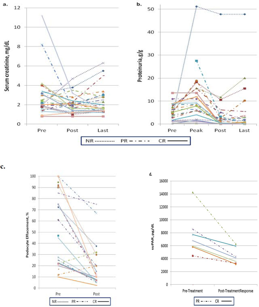
Methods: We conducted a retrospective study of 25 adults with posttransplantation FSGS. We investigated the relationship between suPAR levels and podocyte changes and the impact of therapy on podocyte structure. We assessed response to therapy by improvement in proteinuria, allograft function, and resolution of histologic changes.
Results: A median (interquartile range) of 15 (10-23) plasmapheresis sessions was administered; 13 of the subjects also received rituximab. Median pretreatment suPAR levels were higher among those with severe (≥75%) versus those with mild (≤25%) podocyte foot process effacement (13,030 vs. 4806 pg/mL; P=0.02). Overall, mean±SD of proteinuria improved from 5.1±3.8 to 2.1±2.8 mg/dL (P=0.003), mean podocyte effacement decreased from 57%±33% to 22%±22% (P=0.0001), estimated glomerular filtration rates increased from median (interquartile range) of 32.9 (20.6-44.2) to 39.3 (28.8-63.4; P<0.0001), and suPAR levels decreased from a median of 6.781 to 4.129 pg/mL (P=0.02) with therapy.
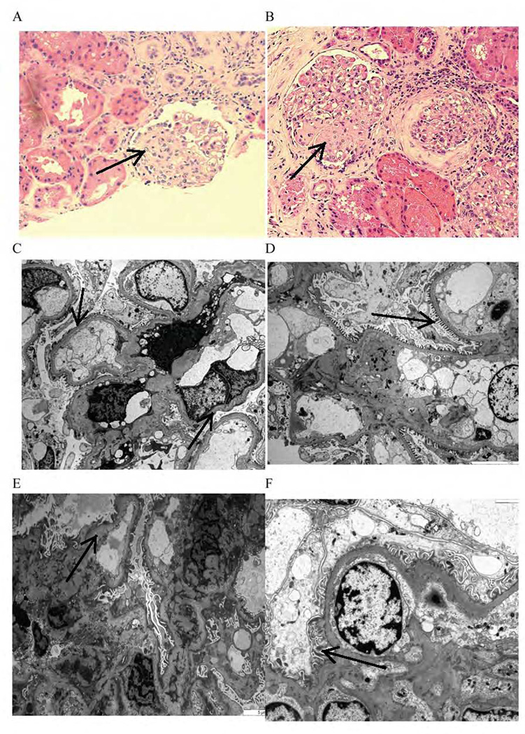
Conclusions: Podocyte effacement is the first pathologic manifestation of FSGS after transplantation. The degree of podocyte effacement correlates with suPAR levels at time of diagnosis. Response to therapy results in significant reduction of suPAR levels and complete or significant improvement of podocyte effacement.
Conflict of interest statement
Figures
References
-
- Kitiyakara C, Eggers P, Kopp JB. Twenty-one-year trend in ESRD due to focal segmental glomerulosclerosis in the United States. Am J Kidney Dis. 2004;44:815. - PubMed
-
- Hickson LJ, Gera M, Amer H, et al. Kidney transplantation for primary focal segmental glomerulosclerosis: outcomes and response to therapy for recurrence. Transplantation. 2009;87:1232. - PubMed
-
- Abbott KC, Sawyers ES, Oliver JD, 3rd, et al. Graft loss due to recurrent focal segmental glomerulosclerosis in renal transplant recipients in the United States. Am J Kidney Dis. 2001;37:366. - PubMed
-
- Savin VJ, Sharma R, Sharma M, et al. Circulating factor associated with increased glomerular permeability to albumin in recurrent focal segmental glomerulosclerosis. N Engl J Med. 1996;334:878. - PubMed
Publication types
MeSH terms
Substances
Grants and funding
LinkOut - more resources
Full Text Sources
Other Literature Sources
Medical
