

Characterization of spatially varying aberrations for wide field-of-view microscopy
- PMID: 23842300
- PMCID: PMC3724395
- DOI: 10.1364/OE.21.015131
Characterization of spatially varying aberrations for wide field-of-view microscopy
Abstract
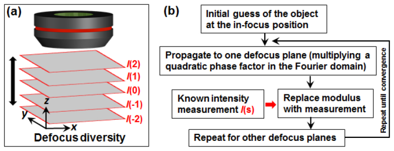
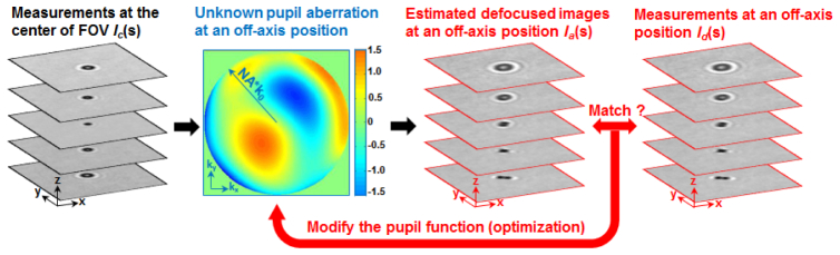
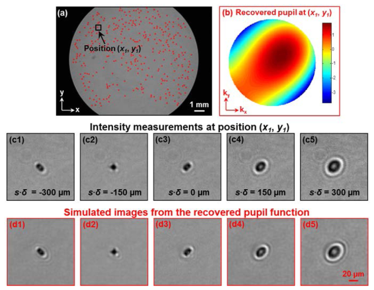
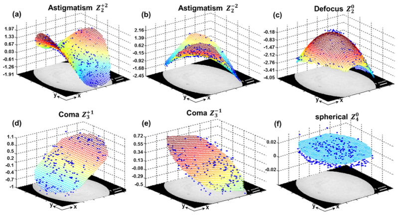
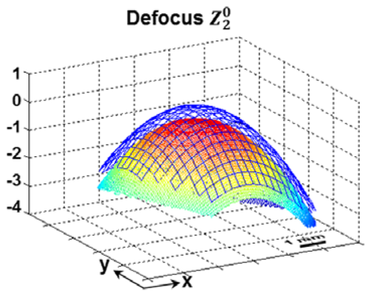
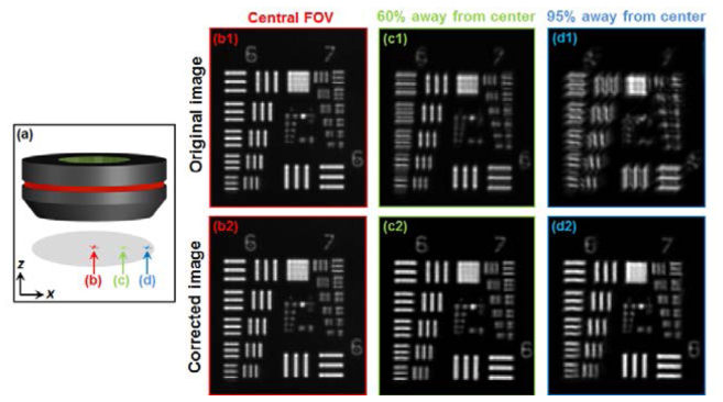
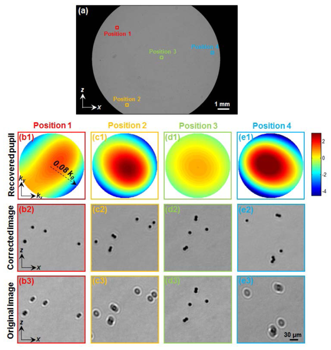
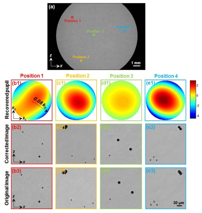
We describe a simple and robust approach for characterizing the spatially varying pupil aberrations of microscopy systems. In our demonstration with a standard microscope, we derive the location-dependent pupil transfer functions by first capturing multiple intensity images at different defocus settings. Next, a generalized pattern search algorithm is applied to recover the complex pupil functions at ~350 different spatial locations over the entire field-of-view. Parameter fitting transforms these pupil functions into accurate 2D aberration maps. We further demonstrate how these aberration maps can be applied in a phase-retrieval based microscopy setup to compensate for spatially varying aberrations and to achieve diffraction-limited performance over the entire field-of-view. We believe that this easy-to-use spatially-varying pupil characterization method may facilitate new optical imaging strategies for a variety of wide field-of-view imaging platforms.
Figures
Similar articles
-
Embedded pupil function recovery for Fourier ptychographic microscopy.Opt Express. 2014 Mar 10;22(5):4960-72. doi: 10.1364/OE.22.004960. Opt Express. 2014. PMID: 24663835 Free PMC article.
-
Tandem aberration correction optics (TACO) in wide-field structured illumination microscopy.Biomed Opt Express. 2023 Nov 20;14(12):6381-6396. doi: 10.1364/BOE.503801. eCollection 2023 Dec 1. Biomed Opt Express. 2023. PMID: 38420301 Free PMC article.
-
Computational aberration compensation by coded-aperture-based correction of aberration obtained from optical Fourier coding and blur estimation.Optica. 2019 May;6(5):647-661. doi: 10.1364/optica.6.000647. Epub 2019 May 10. Optica. 2019. PMID: 33134437 Free PMC article.
-
Phase-retrieved pupil functions in wide-field fluorescence microscopy.J Microsc. 2004 Oct;216(Pt 1):32-48. doi: 10.1111/j.0022-2720.2004.01393.x. J Microsc. 2004. PMID: 15369481
-
Modeling human eye aberrations and their compensation for high-resolution retinal imaging.Optom Vis Sci. 1998 Nov;75(11):827-39. doi: 10.1097/00006324-199811000-00025. Optom Vis Sci. 1998. PMID: 9848838 Review.
Cited by
-
Adaptive correction method of hybrid aberrations in Fourier ptychographic microscopy.J Biomed Opt. 2023 Mar;28(3):036006. doi: 10.1117/1.JBO.28.3.036006. Epub 2023 Mar 13. J Biomed Opt. 2023. PMID: 36923986 Free PMC article.
-
Optical imaging featuring both long working distance and high spatial resolution by correcting the aberration of a large aperture lens.Sci Rep. 2018 Jun 15;8(1):9165. doi: 10.1038/s41598-018-27289-1. Sci Rep. 2018. PMID: 29907794 Free PMC article.
-
Wide-field, high-resolution Fourier ptychographic microscopy.Nat Photonics. 2013 Sep 1;7(9):739-745. doi: 10.1038/nphoton.2013.187. Nat Photonics. 2013. PMID: 25243016 Free PMC article.
-
Ring deconvolution microscopy: exploiting symmetry for efficient spatially varying aberration correction.Nat Methods. 2025 Jun;22(6):1311-1320. doi: 10.1038/s41592-025-02684-5. Epub 2025 Apr 29. Nat Methods. 2025. PMID: 40301622 Free PMC article.
-
Technologies for imaging neural activity in large volumes.Nat Neurosci. 2016 Aug 26;19(9):1154-64. doi: 10.1038/nn.4358. Nat Neurosci. 2016. PMID: 27571194 Free PMC article. Review.
References
-
- H. Gross, W. Singer, M. Totzeck, F. Blechinger, and B. Achtner, Handbook of Optical Systems (Wiley Online Library, 2005), Vol. 2.
-
- F. Berny and S. Slansky, “Wavefront determination resulting from Foucault test as applied to the human eye and visual instruments,” in Optical Instruments and Techniques (Oriel, 1969), pp. 375–386.
Publication types
Grants and funding
LinkOut - more resources
Full Text Sources
Other Literature Sources
