. 2013 Jul 18;499(7458):350-4.
doi: 10.1038/nature12377. Epub 2013 Jul 10.

Structure and function of the Salmonella Typhi chimaeric A(2)B(5) typhoid toxin

Affiliations

Structure and function of the Salmonella Typhi chimaeric A(2)B(5) typhoid toxin

Jeongmin Song et al. Nature. .

Abstract

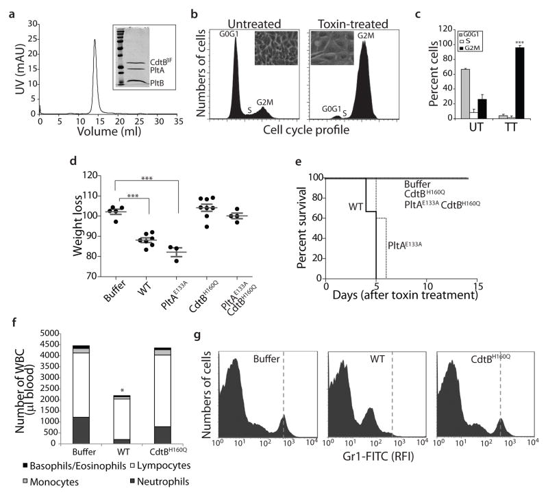
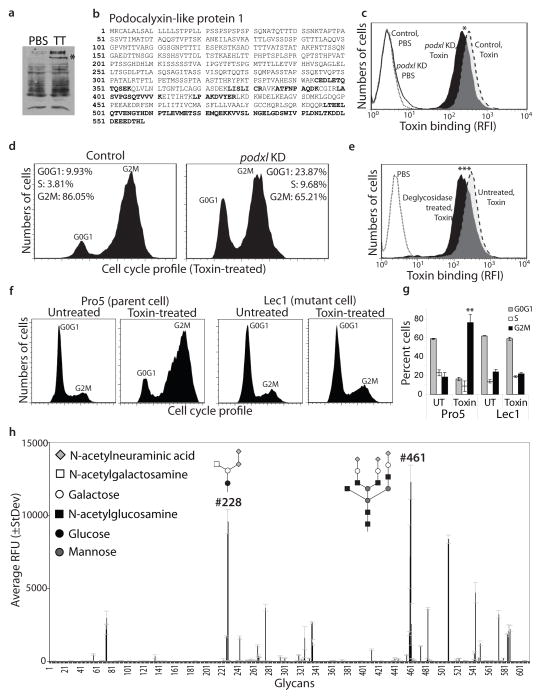
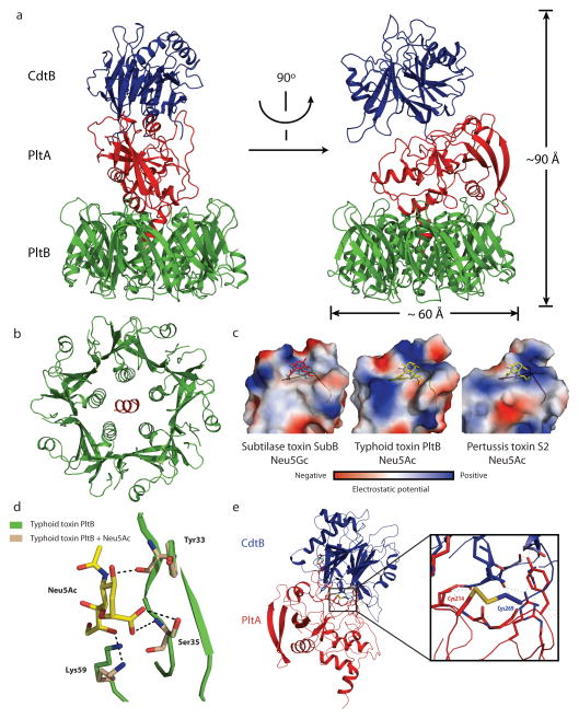
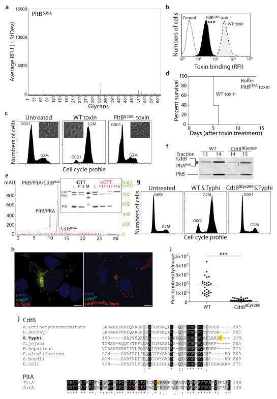
Salmonella enterica serovar Typhi (S. Typhi) differs from most other salmonellae in that it causes a life-threatening systemic infection known as typhoid fever. The molecular bases for its unique clinical presentation are unknown. Here we find that the systemic administration of typhoid toxin, a unique virulence factor of S. Typhi, reproduces many of the acute symptoms of typhoid fever in an animal model. We identify specific carbohydrate moieties on specific surface glycoproteins that serve as receptors for typhoid toxin, which explains its broad cell target specificity. We present the atomic structure of typhoid toxin, which shows an unprecedented A2B5 organization with two covalently linked A subunits non-covalently associated to a pentameric B subunit. The structure provides insight into the toxin's receptor-binding specificity and delivery mechanisms and reveals how the activities of two powerful toxins have been co-opted into a single, unique toxin that can induce many of the symptoms characteristic of typhoid fever. These findings may lead to the development of potentially life-saving therapeutics against typhoid fever.


Conflict of interest statement

The authors declare no financial competing interests. Readers are welcome to comment on the on line version of the manuscript.

Figures

Figure 1
Figure 1
Systemic administration of typhoid toxin causes symptoms observed during the acute phase of typhoid fever. a, Chromatographic profile of the typhoid toxin holotoxin used in the biological assays. The inset shows a Coomassie blue stained SDS-PAGE analysis of the peak fraction. b and c, Typhoid toxin induces cell cycle arrest in cultured cells. Human intestinal epithelial Henle-407 cells were left untreated, or treated with purified typhoid toxin and then analyzed by flow cytometry. The insets show representative light microscope images of mock or toxin treated cells and is representative of at least three independent experiments. c, Averages of cell cycle profiles from at least 3 independent experiments. Bar represents average ± standard deviation. ***, P < 0.001, compared to the number of cells in G2M of the control untreated group. UT: untreated; TT: toxin treated. d, Weight loss 5 days after intravenous administration of different typhoid toxin preparations. Lines are the mean ± standard error of the mean and represent the weight relative to the values before treatment. ***, P < 0.0001. e, Survival of animals receiving different typhoid toxin preparations. n= 3–5 animals per group. f and g, Circulating white blood cells were counted in a hematology analyzer after the indicated treatments (*, P < 0.05) (f). Alternatively, the number of neutrophils (vertical dashed line) in peripheral blood of animals treated as indicated was determined by flow cytometry. RFI, relative fluorescence intensity.
Figure 2
Figure 2
Typhoid toxin recognizes terminally sialylated glycans on surface glycoproteins. a and b, Henle-407 cell surface proteins were biotinlyated, co-immunoprecipitated with purified typhoid toxin (TT), and analyzed by SDS-PAGE (a) and LC-MS/MS. The peptides from Podocalyxin like protein 1 (PODXL) (indicated by an asterix in a) identified by LC-MS/MS are indicated in bold (b). c and d, PODXL-depleted (by an specifically targeted siRNA) and control cells were treated with fluorescently-labeled typhoid toxin and toxin binding was evaluated by flow cytometry (c) (* P=0.024 for three independent determinations). Alternatively, siRNA-depleted and control cells were treated with typhoid toxin and toxicity was evaluated by cell cycle analysis. e, Henle-407 cells were treated with a mixture of glycosidases and the ability of treated and control cells to bind fluorescently-labeled toxin was subsequently evaluated by flow cytometry (***, P < 0.001 from three independent experiments). f and g, The N-acetylglucosaminyltransferase I-deficient (Lec1) and its parent (Pro5) cell lines were treated with typhoid toxin and toxicity was evaluated by cell cycle analysis (f). The quantification of the cell cycle profiles is shown in (g). Bar represents average ± standard deviation of at least three independent determinations. **, P < 0.01, compared to the number of Pro5 cells in G2M. h, Glycan array analysis of typhoid toxin binding. Values represent the average relative fluorescence unit (RFU). The X axis depicts the glycan numbers. The structure of the most relevant glycans is shown.
Figure 3
Figure 3
The crystal structure of typhoid toxin depicts a unique architecture. a, Two views of the overall structure of the typhoid holotoxin complex shown as a ribbon cartoon and related by 90° rotation about a vertical axis. CdtB, PltA and PltB are shown in blue, red, and green, respectively. b, Bottom view of the channel formed by the PltB pentamer (in green), depicting the PltA C-terminal α-helix (in red) within it. c, Surface charge distribution of the predicted sugar-binding pockets of different B subunit homologs of the indicated AB5 toxins (SubB for Subtilase and S2 for Pertussis toxins). A highly conserved serine residue critical for sugar binding is indicated within the sugar-binding pocket. The sugars N-glycolylneuraminic acid (within SubB) and N-acetylneuraminic acid (within typhoid and pertussis toxins) are shown. d, Molecular modeling of N-acetylneuraminic acid within the typhoid toxin binding pocket. Critical residues engaged in this interaction are shown. e, Atomic interface between CdtB and PltA. The inset shows a detailed view of a critical disulfide bond between PltA Cys214 and CdtB Cys269.
Figure 4
Figure 4
Structure-function analysis of typhoid toxin. a–d, Fluorescently-labeled typhoid toxin containing PltBSer35A was tested for its binding to glycans (a) and to cultured cells (b) (see Fig. 2 for details) (***, P < 0.001 from at least three independent determinations). Alternatively, toxicity was assayed by flow cytometric cell cycle analysis of toxin-treated cells (at least three independent experiments) (c), or by systemic administration to mice (n=3 to 5 mice) (d). e–i, The typhoid toxin complex was analyzed by ion exchange chromatography before (blue) and after (red) treatment with DTT (L: loading control; M: molecular weight markers; F: chromatographic fraction) (e). Inset shows SDS-PAGE analyzes of the indicated fractions (e). f, A toxin preparation obtained from a bacterial strain expressing CdtBΔcys269 was analyzed by gel filtration chromatography and compared to wild-type toxin (the experiment was repeated two times). While wild-type holotoxin eluted in fractions 13 and 14, toxin obtained from a bacterial strain encoding CdtBΔcys269 eluted in fractions 14 and 15 due to the lack of CdtB. g–i, Henle-407 cells were infected with S. Typhi strains encoding FLAG-tagged CdtB or CdtBΔcys269 and cells were examined for toxicity by flow cytometry (g). Alternatively, cells were fixed, stained with anti FLAG antibody, and the amount of puncta staining, which represent CdtB in typhoid toxin export carriers, were determined by immunofluorescence analysis (h and i). Bar represents average of puncta-associated flurorescence intensity (at least 100 cells were analyzed in three independent experiments).***, P < 0.0001, Scale bar:10 μm. j, ClustalW amino acid sequence comparison of CdtB and PltA homologs. Conserved cysteines are shown in red while unique cysteines are indicagted with a yellow shade.

Comment in

  • Bacteriology: toxins in tandem.
    Stebbins CE. Stebbins CE. Nature. 2013 Jul 18;499(7458):293. doi: 10.1038/nature12409. Epub 2013 Jul 10. Nature. 2013. PMID: 23842489 No abstract available.

Similar articles

Cited by

References

    1. Parry C, Hien TT, Dougan G, White N, Farrar J. Typhoid fever. N Engl J Med. 2002;347:1770–1782. - PubMed
    1. Parkhill J, et al. Complete genome sequence of a multiple drug resistant Salmonella enterica serovar Typhi CT18. Nature. 2001;413:848–852. - PubMed
    1. Butler T. Treatment of typhoid fever in the 21st century: promises and shortcomings. Clin Microbiol Infect. 2011;17:959–963. - PubMed
    1. Crump J, Mintz E. Global trends in typhoid and paratyphoid Fever. Clin Infect Dis. 2010;50:241–246. - PMC - PubMed
    1. Sabbagh S, Forest C, Lepage C, Leclerc J, Daigle F. So similar, yet so different: uncovering distinctive features in the genomes of Salmonella enterica serovars Typhimurium and Typhi. FEMS Microbiol Lett. 2010;305:1–13. - PubMed

Publication types

Associated data