

An experimental protocol for in vivo imaging of neuronal structural plasticity with 2-photon microscopy in mice
- PMID: 23842538
- PMCID: PMC3716956
- DOI: 10.1186/2040-7378-5-9
An experimental protocol for in vivo imaging of neuronal structural plasticity with 2-photon microscopy in mice
Abstract
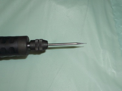
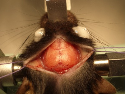
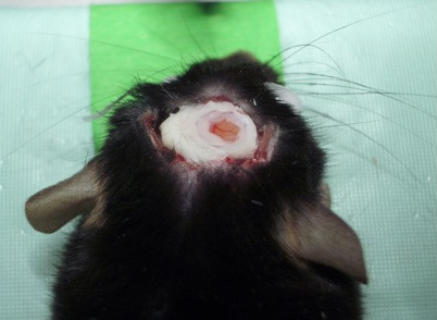
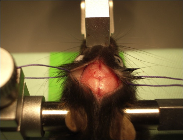
Introduction: Structural plasticity with synapse formation and elimination is a key component of memory capacity and may be critical for functional recovery after brain injury. Here we describe in detail two surgical techniques to create a cranial window in mice and show crucial points in the procedure for long-term repeated in vivo imaging of synaptic structural plasticity in the mouse neocortex.
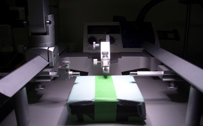
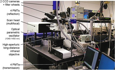
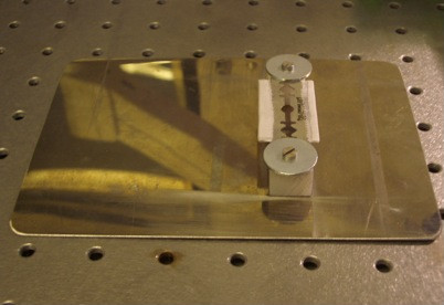
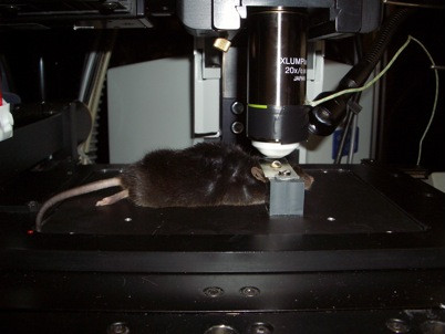
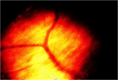
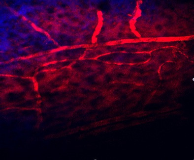
Methods: Transgenic Thy1-YFP(H) mice expressing yellow-fluorescent protein (YFP) in layer-5 pyramidal neurons were prepared under anesthesia for in vivo imaging of dendritic spines in the parietal cortex either with an open-skull glass or thinned skull window. After a recovery period of 14 days, imaging sessions of 45-60 min in duration were started under fluothane anesthesia. To reduce respiration-induced movement artifacts, the skull was glued to a stainless steel plate fixed to metal base. The animals were set under a two-photon microscope with multifocal scanhead splitter (TriMScope, LaVision BioTec) and the Ti-sapphire laser was tuned to the optimal excitation wavelength for YFP (890 nm). Images were acquired by using a 20×, 0.95 NA, water-immersion objective (Olympus) in imaging depth of 100-200 μm from the pial surface. Two-dimensional projections of three-dimensional image stacks containing dendritic segments of interest were saved for further analysis. At the end of the last imaging session, the mice were decapitated and the brains removed for histological analysis.
Results: Repeated in vivo imaging of dendritic spines of the layer-5 pyramidal neurons was successful using both open-skull glass and thinned skull windows. Both window techniques were associated with low phototoxicity after repeated sessions of imaging.
Conclusions: Repeated imaging of dendritic spines in vivo allows monitoring of long-term structural dynamics of synapses. When carefully controlled for influence of repeated anesthesia and phototoxicity, the method will be suitable to study changes in synaptic structural plasticity after brain injury.
Figures


References
LinkOut - more resources
Full Text Sources
Other Literature Sources
Miscellaneous
