

Review
doi: 10.1007/128_2013_464.
UDP-GlcNAc 2-Epimerase/ManNAc Kinase (GNE): A Master Regulator of Sialic Acid Synthesis
Affiliations
- PMID: 23842869
- PMCID: PMC4161665
- DOI: 10.1007/128_2013_464
Item in Clipboard
Review
UDP-GlcNAc 2-Epimerase/ManNAc Kinase (GNE): A Master Regulator of Sialic Acid Synthesis
Top Curr Chem.
2015.
Abstract
UDP-N-acetylglucosamine 2-epimerase/N-acetylmannosamine kinase is the key enzyme of sialic acid biosynthesis in vertebrates. It catalyzes the first two steps of the cytosolic formation of CMP-N-acetylneuraminic acid from UDP-N-acetylglucosamine. In this review we give an overview of structure, biochemistry, and genetics of the bifunctional enzyme and its complex regulation. Furthermore, we will focus on diseases related to UDP-N-acetylglucosamine 2-epimerase/N-acetylmannosamine kinase.
Figures
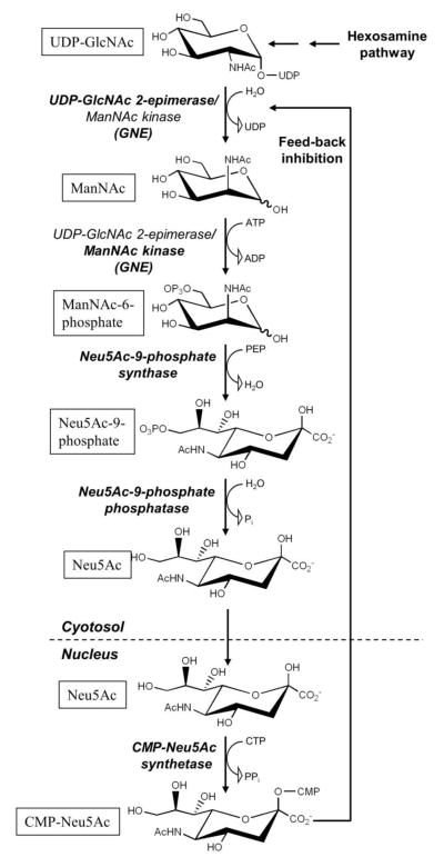
GNE catalyzes the first two steps of this pathway. The respective enzymatic reactions are indicated in bold. Note, that UDP-GlcNAc is supplied from fructose-6-phosphate by the hexosamine pathway, and that CMP-Neu5Ac is formed in the nucleus. Feed-back inhibition of the UDP-GlcNAc 2-epimerase activity of GNE by CMP-Neu5Ac is further indicated.
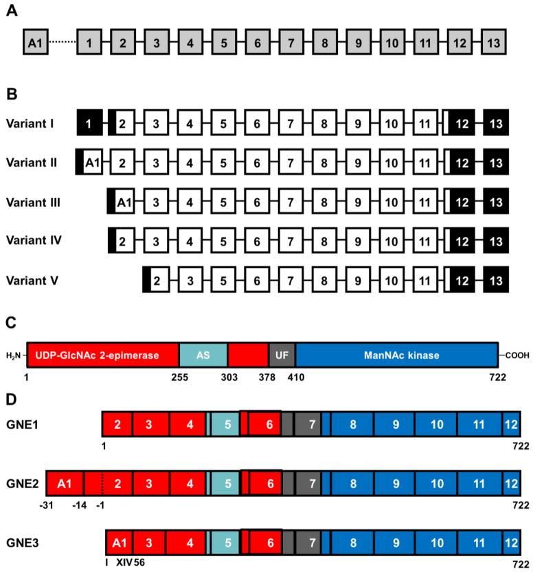
(A) Schematic exon structure of the human GNE gene. (B) Predicted GNE mRNAs by alternative splicing. (C) Protein domain structure of GNE. (D) Schematic structure of the GNE isoforms with proven enzymatic activity (for details see text).
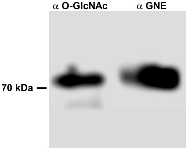
GNE was immunoprecipitated from rat liver by a GNE-specific antibody and Western-blotted. Immunodetection was performed with O-GlcNAc- and GNE-specific antibodies, indicating O-GlcNAcylation of the GNE protein.
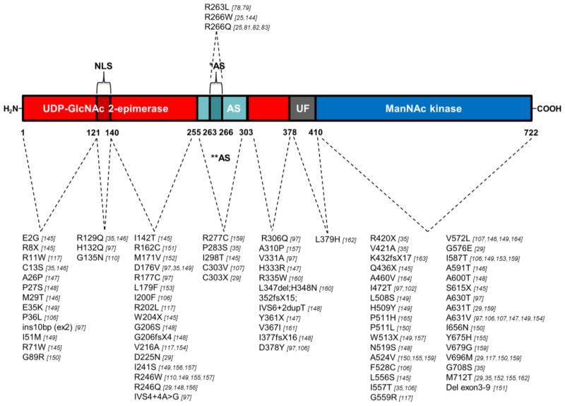
Locations and characteristics of all reported human GNE mutations within the functional domains of the GNE protein (as of April 2011). Mutations associated with sialuria are printed above the protein cartoon, and HIBM-associated mutations below, including their references. Red bar, UDP-GlcNAc 2-epimerase domain (GNE, Ep); dark red bar, the putative GNE nuclear export signal (Ep (N)); green bar, experimental allosteric region based on in vitro studies (**AS); dark green bar, allosteric site based on human sialuria mutations (*AS); gray bar, region of unknown function (UF); blue bar, ManNAc kinase domain (MNK, Kin). Note, that references [–165] were only given in this figure.
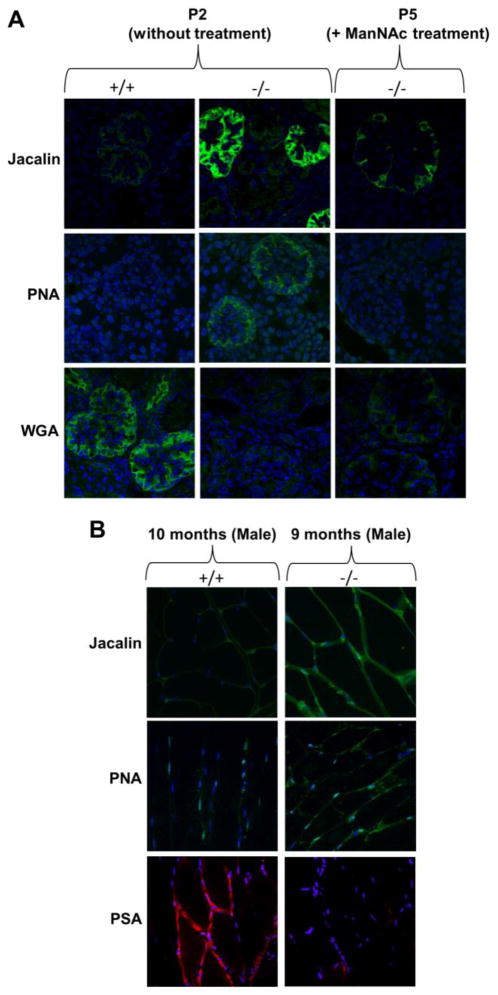
Representative images of kidney glomeruli and quadriceps muscle cells in paraffin embedded mouse slides stained with FITC-labeled lectins (green) Jacalin (jackfruit agglutinin) and PNA (peanut agglutinin), which both bind predominantly to terminal galactose residues residues, and WGA (wheat germ agglutinin), which predominantly recognizes terminal Neu5Ac and GlcNAc. Muscle samples were also treated with antibodies to polysialic acid (PSA) (red). All slides were counterstained with the nuclear dye DAPI (blue). Confocal imaging intensity settings were the same across all ages and genotypes for each lectin or antibody. (A) Glomeruli from Gne M712T knock-in HIBM pups at postnatal day 2 (P2) showed hyposialylation in mutant (−/ −) compared to wild type (+/+) kidneys, as demonstrated by increased Jacalin and PNA signals and decreased WGA signal in −/ − glomeruli compared to +/+. After ManNAc treatment at P5, −/ − glomeruli show decreased Jacalin and PNA signals a more intense WGA signal compared to −/ − glomeruli at P2 without treatment, suggestive of increased sialylation after ManNAc treatment. (B) Quadriceps muscles from adult male mice (+/+, 10 months; −/ −, 9 months old) showed increased Jacalin and PNA signals and decreased PSA staining in −/ − compared to +/+ tissues, indicating hyposialylation of muscle glycans in adult HIBM mice mimicking the human disorder.
References
-
- Luchansky SJ, Yarema KJ, Takahashi S, Bertozzi CR. GlcNAc 2-epimerase can serve a catabolic role in sialic acid metabolism. J Biol Chem. 2003;278:8035–8042. - PubMed
-
- Hinderlich S, Berger M, Keppler OT, Pawlita M, Reutter W. Biosynthesis of N-acetylneuraminic acid in cells lacking UDP-N-acetylglucosamine 2-epimerase/N-acetylmannosamine kinase. Biol Chem. 2001;382:291–297. - PubMed
-
- Schauer R, Wember M. Isolation and characterization of sialate lyase from pig kidney. Biol Chem Hoppe Seyler. 1996;377:293–299. - PubMed
Publication types
MeSH terms
Substances
Grants and funding
LinkOut - more resources
Full Text Sources
Other Literature Sources
